FC Flow cytometry


PAX5 (D19F8) XP ® Rabbit mAb (Alexa Fluor ® 647 Conjugate) - 11852 from Cell Signaling Technology

Host Rabbit
Clonality Monoclonal
Clone D19F8
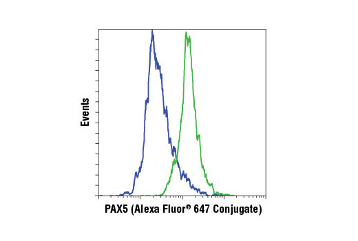
Flow cytometric analysis of Jurkat (blue) and Ramos (green) cells using PAX5 (D19F8) XP® Rabbit mAb (Alexa Fluor® 647 Conjugate).

Phospho-p44/42 MAPK (Erk1/2) (Thr202/Tyr204) (D13.14.4E) XP ® Rabbit mAb (Alexa Fluor ® 647 Conjugate) - 4284 from Cell Signaling Technology

Host Rabbit
Clonality Monoclonal
Clone D13.14.4E
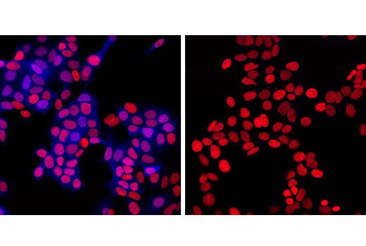
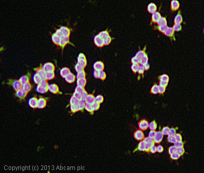
Confocal immunofluorescent analysis of MCF7 cells, insulin-treated (100 nM, 10 minutes; left) or treated with U0126 #9903 (10 uM, 2 hours; right), using Phospho-p44/42 MAPK (Erk1/2) (Thr202/Tyr204) (D13.14.4E) XP® Rabbit mAb (Alexa Fluor® 647 Conjugate) (blue pseudocolor). Red = Propidium Iodide (PI)/RNase Staining Solution #4087.

Phospho-S6 Ribosomal Protein (Ser240/244) (61H9) Rabbit mAb (Biotinylated) - 4278 from Cell Signaling Technology

Host Rabbit
Clonality Monoclonal
Clone 61H9
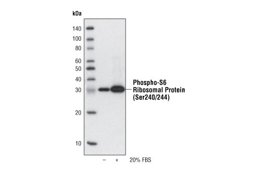
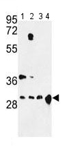
Western blot analysis of extracts from NIH/3T3 cells, untreated or treated with 20% fetal bovine serum (FBS), using Phospho-S6 Ribosomal Protein (Ser240/244) (61H9) Rabbit mAb (Biotinylated) and developed with Streptavidin-HRP #3999.

Ku-70/Ku-86 (162) - sc-53632 from Santa Cruz Biotechnology

Host Mouse
Clonality Monoclonal
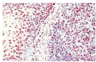
Ku-70/Ku-86 (162): sc-53632. Immunoperoxidase staining of formalin-fixed, paraffin-embedded human tonsil tissue showing nuclear localization.

Tyrosinase (M-19) - sc-7834 from Santa Cruz Biotechnology

Host Goat
Clonality Polyclonal
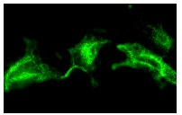
Tyrosinase (M-19): sc-7834. Immunofluorescence staining of methanol-fixed B16-FO cells showing cytoplasmic staining.

H2-Kb (DaB1) - sc-65382 from Santa Cruz Biotechnology

Host Mouse
Clonality Monoclonal

Integrin αM (Bear-1) - sc-52686 from Santa Cruz Biotechnology

Host Mouse
Clonality Monoclonal
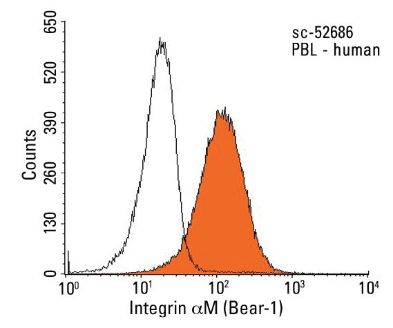
Integrin αM (Bear-1): sc-52686. Indirect FCM analysis of human peripheral blood leukocytes stained with Integrin αM (Bear-1), followed by PE-conjugated goat anti-mouse IgG: sc-3738. Black line histogram represents the isotype control, normal mouse IgG<sub>1</sub>: sc-3877.

MAdCAM-1 (17F5) - sc-59790 from Santa Cruz Biotechnology

Host Mouse
Clonality Monoclonal

Anti-14-3-3 alpha + beta antibody [Y62] - ab32560 from Abcam

Prices $357.00
Sizes 100 µl
Host Rabbit
Clonality Monoclonal
Clone Y62
Anti-14-3-3 alpha + beta antibody [Y62] (ab32560) at 1/1000 dilution + Hela cell lysate.

Anti-14-3-3 antibody [E58] - ab32377 from Abcam

Prices $379.00
Sizes 100 µl
Host Rabbit
Clonality Monoclonal
Clone E58
Anti-14-3-3 antibody [E58] (ab32377) at 1/1000 dilution + Hela cell lysate.

Anti-14-3-3 epsilon antibody [EPR3918] - ab92311 from Abcam

Prices $391.00
Sizes 100 µl
Host Rabbit
Clonality Monoclonal
Clone EPR3918
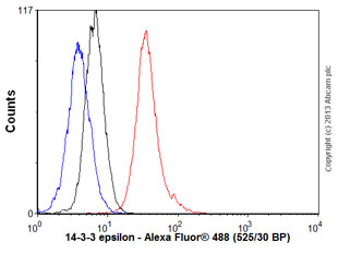
Overlay histogram showing SH-SY5Y cells stained with ab92311 (red line). The cells were fixed with 80% methanol (5 min) and then permeabilized with 0.1% PBS-Tween for 20 min. The cells were then incubated in 1x PBS / 10% normal goat serum / 0.3M glycine to block non-specific protein-protein interactions followed by the antibody (ab92311, 1/1000 dilution) for 30 min at 22°C. The secondary antibody used was Alexa Fluor® 488 goat anti-rabbit IgG (H&L) (ab150077) at 1/2000 dilution for 30 min at 22°C. Isotype control antibody (black line) was rabbit IgG (monoclonal) (0.1μg/1x106 cells) used under the same conditions. Unlabelled sample (blue line) was also used as a control. Acquisition of >5,000 events were collected using a 20mW Argon ion laser (488nm) and 525/30 bandpass filter. This antibody gave a positive signal in SH-SY5Y cells fixed with 4% paraformaldehyde (10 min)/permeabilized with 0.1% PBS-Tween for 20 min used under the same conditions.

Anti-14-3-3 epsilon antibody [EPR3919] - ab108210 from Abcam

Host Rabbit
Clonality Monoclonal
Clone EPR3919
All lanes : Anti-14-3-3 epsilon antibody [EPR3919] (ab108210)Lane 1 : Fetal brain lysateLane 2 : SW480 cell lysateLane 3 : NIH/3T3 cell lysateLysates/proteins at 10 µg per lane.SecondaryHRP labelled goat anti-rabbit at 1/2000 dilution

Anti-14-3-3 gamma antibody - ab174491 from Abcam

Prices $384.00
Sizes 400 µl
Host Rabbit
Clonality Polyclonal
Anti-14-3-3 gamma antibody (ab174491) at 1/50 dilution + Mouse cerebellum tissue lysate at 35 µg

Anti-14-3-3 gamma antibody [EPR9655(B)] - ab137106 from Abcam

Prices $379.00
Sizes 100 µl
Host Rabbit
Clonality Monoclonal
Clone EPR9655(B)
All lanes : Anti-14-3-3 gamma antibody [EPR9655(B)] (ab137106) at 1/1000 dilutionLane 1 : HeLa cell lysateLane 2 : A431 cell lysateLane 3 : 293T cell lysateLane 4 : K562 cell lysateLysates/proteins at 10 µg per lane.SecondaryHRP-labelled Goat anti-Rabbit at 1/2000 dilution

Anti-14-3-3 Tau antibody [3B9] - ab10439 from Abcam

Prices $391.00
Sizes 100 µg
Host Mouse
Clonality Monoclonal
Clone 3B9
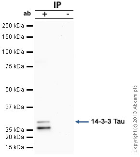
14-3-3 Tau was immunoprecipitated using 0.5mg Hela whole cell extract, 5µg of Mouse monoclonal to 14-3-3 Tau and 50µl of protein G magnetic beads (+). No antibody was added to the control (-).The antibody was incubated under agitation with Protein G beads for 10min, Hela whole cell extract lysate diluted in RIPA buffer was added to each sample and incubated for a further 10min under agitation.Proteins were eluted by addition of 40µl SDS loading buffer and incubated for 10min at 70°C; 10µl of each sample was separated on a SDS PAGE gel, transferred to a nitrocellulose membrane, blocked with 5% BSA and probed with ab10439.Secondary: Goat polyclonal to mouse IgG light chain specific (HRP) at 1/20,000 dilution.Band: 31kDa, non specific band - 26kDa: We are unsure as to the identity of this extra band; 14-3-3 Tau

Anti-14-3-3 Theta + Tau antibody [EPR5123] - ab124909 from Abcam

Prices $313.00
Sizes 100 µl
Host Rabbit
Clonality Monoclonal
Clone EPR5123
All lanes : Anti-14-3-3 Theta + Tau antibody [EPR5123] (ab124909) at 1/10000 dilutionLane 1 : HeLa cell lysateLane 2 : 293T cell lysateLane 3 : A549 cell lysateLane 4 : A431 cell lysateLysates/proteins at 10 µg per lane.SecondaryHRP labelled goat anti-rabbit at 1/2000 dilution

Anti-14-3-3 zeta antibody - ab135823 from Abcam

Prices $384.00
Sizes 400 µl
Host Rabbit
Clonality Polyclonal
All lanes : Anti-14-3-3 zeta antibody (ab135823) at 1/100 dilutionLane 1 : MDA-MB-231 cell lysateLane 2 : A2058 cell lysateLane 3 : Mouse cerebellum tissue lysateLane 4 : Mouse brain tissue lysateLysates/proteins at 35 µg per lane.

Anti-2 Cys Peroxiredoxin antibody [6E5] - ab16765 from Abcam

Prices $380.00
Sizes 100 µl
Host Mouse
Clonality Monoclonal
Clone 6E5
ICC/IF image of ab16765 stained HCT116 cells. The cells were 100% methanol fixed (5 min) and then incubated in 1%BSA / 10% normal goat serum / 0.3M glycine in 0.1% PBS-Tween for 1h to permeabilise the cells and block non-specific protein-protein interactions. The cells were then incubated with the antibody (ab16765, 10µg/ml) overnight at +4°C. The secondary antibody (green) was ab96879, DyLight® 488 goat anti-mouse IgG (H+L) used at a 1/250 dilution for 1h. Alexa Fluor® 594 WGA was used to label plasma membranes (red) at a 1/200 dilution for 1h. DAPI was used to stain the cell nuclei (blue) at a concentration of 1.43µM

Anti-2 Hydroxy phytanoyl CoA lyase antibody [EPR16812] - ab197025 from Abcam

Prices $357.00
Sizes 100 µl
Host Rabbit
Clonality Monoclonal
Clone EPR16812
All lanes : Anti-2 Hydroxy phytanoyl CoA lyase antibody [EPR16812] (ab197025) at 1/1000 dilutionLane 1 : HeLa (Human epithelial cells from cervix adenocarcinoma) whole cell lysateLane 2 : 293T (Human epithelial cells from embryonic kidney) whole cell lysateLysates/proteins at 20 µg per lane.SecondaryGoat Anti-Rabbit IgG, (H+L), Peroxidase conjugated at 1/1000 dilution

Anti-200 kD Neurofilament Heavy antibody [NF-01] - ab7795 from Abcam

Prices $395.00
Sizes 100 µg
Host Mouse
Clonality Monoclonal
Clone NF-01
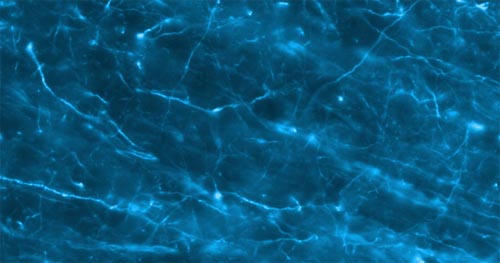
ab7795 at a dilution of 1/1000, staining 200 kD Neurofilament Heavy  (Alexa 488 secondary at 1/2000) in rat brain tissue (30µm thick coronal sections) on free floating IHC (see protocol link for detailed description). Image taken with a 40x objective. No labeling observed following omission of primary antibody.Sections were viewed using an Axioplan 2 Imaging microscope (Imaging Associates) fitted with 10x, 20x and 40x Plan-Neofluorobjectives (Zeiss, Germany) and images were taken using a AxioCam Hrm digital camera (Zeiss, Germany) and AxioVision software (Imaging Associates).

Anti-200 kD Neurofilament Heavy antibody [NF421] - ab187374 from Abcam

Prices $378.00
Sizes 500 µl
Host Mouse
Clonality Monoclonal
Clone NF421
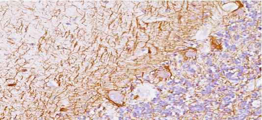
Immunohistochemical analysis of paraffin embedded Human cerebellum tissue labeling Neurofilament with ab187374 at 1 µg/ml.

Anti-3-Nitrotyrosine antibody [7A12AF6] - ab110282 from Abcam

Prices $390.00
Sizes 100 µg
Host Mouse
Clonality Monoclonal
Clone 7A12AF6
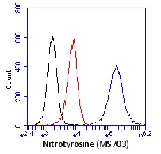
HL-60 cells were stained with 1 µg/ml ab110282 following treatment with 2 mM peroxynitrite (blue) or vehicle control (red). No primary antibody control is shown in black. Peroxynitrite modifies tyrosine residues to 3-nitrotyrosine.

Anti-3-Nitrotyrosine antibody [7A12AF6] (Alexa Fluor® 488) - ab157402 from Abcam

Prices $431.00
Sizes 100 µg
Host Mouse
Clonality Monoclonal
Clone 7A12AF6
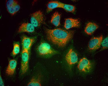
Immunocytochemical analysis of HeLa cells (40x magnification) treated with 1 mM peroxynitrite labeling 3-Nitrotyrosine with ab157402 at 5.0 µg/ml (green); anti-HSP60 (1/3000, ab46798) as red, and DAPI in blue, as a nuclear stain. Secondary antibody used was goat anti-rabbit Alexa594 (1/2000). All blocking and antibodies were diluted in 10% normal goat serum.

Anti-4 Hydroxynonenal antibody [HNEJ-2] - ab48506 from Abcam

Prices $395.00
Sizes 50 µg
Host Mouse
Clonality Monoclonal
Clone HNEJ-2
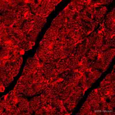
ab48506 at a 1/25 dilution staining 4-Hydroxy-2-Nonenal in mouse heart tissue sections by Immunohistochemistry (paraffin embedded) incubated for 15 hours at +4°C. Fixed with formaldehyde, heat mediated antigen retrieval step performed using citrate buffer. Blocked using 5% serum for 20 minutes at 20°C. Secondary used undiluted polyclonal Goat anti-mouse IgG conjugated to Alexa Fluor 594.See Abreview

Anti-5-methylcytosine (5-mC) antibody [33D3] - ab10805 from Abcam

Prices $397.00
Sizes 50 µg
Host Mouse
Clonality Monoclonal
Clone 33D3
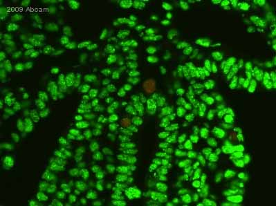
ab10805 staining 5-Methyl Cytidine in pig embryo (15 to 17 days) tissue section by Immunohistochemistry (Formalin/PFA-fixed paraffin-embedded sections). Tissue underwent fixation in paraformaldehyde, heat mediated antigen retrieval in Tris-EDTA buffer, permeabilization in Triton X-100 and blocking in 2% BSA for 10 minutes at 25°C. The primary antibody was diluted, 1/100 (PBS + 2% BSA) and incubated with sample for 1 hour at 25°C. An Alexa Fluor® 488 conjugated donkey polyclonal to mouse at 1/250 dilution, was used as secondary.See Abreview
Search more