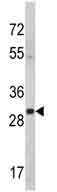
Anti-14-3-3 gamma antibody (ab174491) at 1/50 dilution + Mouse cerebellum tissue lysate at 35 µg
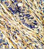
Immunohistochemical analysis of formalin-fixed and paraffin-embedded Human colon carcinoma labeling 14-3-3 gamma with ab174491 at 1/50 dilution followed by peroxidase-conjugated secondary antibody and DAB staining.
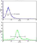
Flow cytometric analysis of widr cells labeling 14-3-3 gamma with ab174491 at 1/10 dilution (bottom histogram) compared to a negative control cell (top histogram). FITC-conjugated goat-anti-rabbit secondary antibodies were used for the analysis.
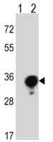
All lanes : Anti-14-3-3 gamma antibody (ab174491) at 1/50 dilutionLane 1 : Non-transfected 293 cell lysateLane 2 : 14-3-3 gamma transfected 293 cell lysateLysates/proteins at 2 µg per lane.