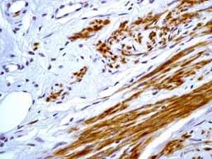
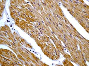
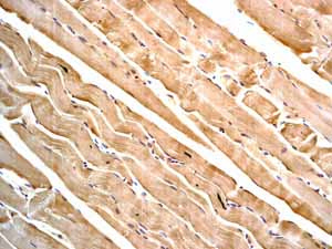
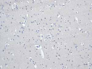
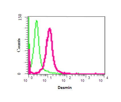
Anti-Desmin antibody [Y66] - Cytoskeleton Marker
| Name | Anti-Desmin antibody [Y66] - Cytoskeleton Marker |
|---|---|
| Supplier | Abcam |
| Catalog | ab32362 |
| Prices | $404.00 |
| Sizes | 100 µl |
| Host | Rabbit |
| Clonality | Monoclonal |
| Isotype | IgG |
| Clone | Y66 |
| Applications | ICC/IF ICC/IF WB IHC-P IHC FC IHC-F |
| Species Reactivities | Mouse, Rat, Guinea Pig, Human |
| Antigen | Synthetic peptide (the amino acid sequence is considered to be commercially sensitive) corresponding to Human Desmin aa 400 to the C-terminus (C terminal) |
| Description | Rabbit Monoclonal |
| Gene | DES |
| Conjugate | Unconjugated |
| Supplier Page | Shop |
Product images
Product References
POMK mutation in a family with congenital muscular dystrophy with merosin - POMK mutation in a family with congenital muscular dystrophy with merosin
von Renesse A, Petkova MV, Lutzkendorf S, Heinemeyer J, Gill E, Hubner C, von Moers A, Stenzel W, Schuelke M. J Med Genet. 2014 Apr;51(4):275-82.
Potential of adipose-derived mesenchymal stem cells and skeletal muscle-derived - Potential of adipose-derived mesenchymal stem cells and skeletal muscle-derived
Ren Y, Wu H, Ma Y, Yuan J, Liang H, Liu D. PLoS One. 2014 Apr 3;9(4):e93583.
Desmoplakin truncations and arrhythmogenic left ventricular cardiomyopathy: - Desmoplakin truncations and arrhythmogenic left ventricular cardiomyopathy:
Lopez-Ayala JM, Gomez-Milanes I, Sanchez Munoz JJ, Ruiz-Espejo F, Ortiz M, Gonzalez-Carrillo J, Lopez-Cuenca D, Oliva-Sandoval MJ, Monserrat L, Valdes M, Gimeno JR. Europace. 2014 Dec;16(12):1838-46.
The Ran GTPase-activating protein (RanGAP1) is critically involved in smooth - The Ran GTPase-activating protein (RanGAP1) is critically involved in smooth
Vorpahl M, Schonhofer-Merl S, Michaelis C, Flotho A, Melchior F, Wessely R. PLoS One. 2014 Jul 2;9(7):e101519.
Angioleiomyomas in the head and neck: A retrospective clinical and - Angioleiomyomas in the head and neck: A retrospective clinical and
Liu Y, Li B, Li L, Liu Y, Wang C, Zha L. Oncol Lett. 2014 Jul;8(1):241-247. Epub 2014 May 8.
SPEG interacts with myotubularin, and its deficiency causes centronuclear - SPEG interacts with myotubularin, and its deficiency causes centronuclear
Agrawal PB, Pierson CR, Joshi M, Liu X, Ravenscroft G, Moghadaszadeh B, Talabere T, Viola M, Swanson LC, Haliloglu G, Talim B, Yau KS, Allcock RJ, Laing NG, Perrella MA, Beggs AH. Am J Hum Genet. 2014 Aug 7;95(2):218-26.
IQGAP1 suppresses TbetaRII-mediated myofibroblastic activation and metastatic - IQGAP1 suppresses TbetaRII-mediated myofibroblastic activation and metastatic
Liu C, Billadeau DD, Abdelhakim H, Leof E, Kaibuchi K, Bernabeu C, Bloom GS, Yang L, Boardman L, Shah VH, Kang N. J Clin Invest. 2013 Mar 1;123(3):1138-56.
Exacerbated neuronal ceroid lipofuscinosis phenotype in Cln1/5 double-knockout - Exacerbated neuronal ceroid lipofuscinosis phenotype in Cln1/5 double-knockout
Blom T, Schmiedt ML, Wong AM, Kyttala A, Soronen J, Jauhiainen M, Tyynela J, Cooper JD, Jalanko A. Dis Model Mech. 2013 Mar;6(2):342-57.
Characterization and inflammatory response of perivascular-resident - Characterization and inflammatory response of perivascular-resident
Zhang F, Zhang J, Neng L, Shi X. J Assoc Res Otolaryngol. 2013 Oct;14(5):635-43.
Single-cell clones of liver cancer stem cells have the potential of - Single-cell clones of liver cancer stem cells have the potential of
Liu H, Zhang W, Jia Y, Yu Q, Grau GE, Peng L, Ran Y, Yang Z, Deng H, Lou J. Cell Death Dis. 2013 Oct 17;4:e857.
![All lanes : Anti-Desmin antibody [Y66] - Cytoskeleton Marker (ab32362) at 1/500000 dilution (purified)Lane 1 : Human skeletal muscle tissue lysateLane 2 : Human fetal heart tissue lysateLane 3 : Human fetal muscle tissue lysateLysates/proteins at 20 µg per lane.SecondaryHRP-conjugated anti-rabbit IgG, specific to the non-reduced form of IgG at 1/1000 dilution](http://www.bioprodhub.com/system/product_images/ab_products/2/sub_2/7854_ab32362-239396-ab32362wb.jpg)
![All lanes : Anti-Desmin antibody [Y66] - Cytoskeleton Marker (ab32362) at 1/500000 dilution (purified)Lane 1 : Mouse heart tissue lysateLane 2 : Rat heart tissue lysateLysates/proteins at 20 µg per lane.SecondaryHRP-conjugated anti-rabbit IgG, specific to the non-reduced form of IgG at 1/1000 dilution](http://www.bioprodhub.com/system/product_images/ab_products/2/sub_2/7855_ab32362-239397-ab32362wb2.jpg)
![All lanes : Anti-Desmin antibody [Y66] - Cytoskeleton Marker (ab32362) at 1/100000 dilutionLane 1 : Guinea pig heart tissue lysateLane 2 : Guinea pig muscle tissue lysateLysates/proteins at 10 µg per lane.SecondaryPeroxidase-conjugated goat anti-rabbit IgG (H+L) at 1/1000 dilution](http://www.bioprodhub.com/system/product_images/ab_products/2/sub_2/7856_ab32362-239398-ab32362wb3.jpg)
![All lanes : Anti-Desmin antibody [Y66] - Cytoskeleton Marker (ab32362) at 1/500 dilution (unpurified)Lane 1 : Human skeletal muscle whole tissue lysateLane 2 : Human skeletal muscle whole tissue lysateLane 3 : Human skeletal muscle whole tissue lysateLane 4 : Human skeletal muscle whole tissue lysateLysates/proteins at 20 µg per lane.SecondaryIRDye® 680-conjugated anti-rabbit at 1/5000 dilutiondeveloped using the ECL techniquePerformed under reducing conditions.](http://www.bioprodhub.com/system/product_images/ab_products/2/sub_2/7857_ab32362-207533-ab32362wb.jpg)
