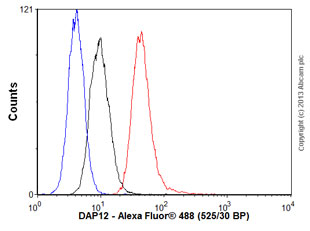
Overlay histogram showing THP1 cells stained with ab124834 (red line). The cells were fixed with 80% methanol (5 min) and then permeabilized with 0.1% PBS-Tween for 20 min. The cells were then incubated in 1x PBS / 10% human serum / 0.3M glycine to block non-specific protein-protein interactions followed by the antibody (ab124834, 1/100 dilution) for 30 min at 22°C. The secondary antibody used was Alexa Fluor® 488 goat anti-rabbit IgG (H&L) (ab150077) at 1/2000 dilution for 30 min at 22°C. Isotype control antibody (black line) was rabbit IgG (monoclonal) (1μg/1x106 cells) used under the same conditions. Unlabelled sample (blue line) was also used as a control. Acquisition of >5,000 events were collected using a 20mW Argon ion laser (488nm) and 525/30 bandpass filter.
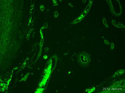
IHC-Fr image of human blood vessels stained with Ab124834 (1/500). The fixed brain section were permeabilise in 0.1% PBS-Triton X for 1h. The sections were incubated with 10% donkey serum for 1 h at 24°C to block non-specific protein-protein interactions. The sections were then incubated with Ab124834 used at 1/500 dilution, overnight at 4°C. The secondary antibody was Alexa Fluor® 488 donkey anti-rabbit used at a 1/1000 dilution for 1h. DAPI was used to stain the cell nuclei (blue) at a concentration of 1.43µM.See Abreview
![All lanes : Anti-DAP12 antibody [EPR5173] (ab124834) at 1/1000 dilutionLane 1 : Peripheral blood leukocytes lysatesLane 2 : Human placenta lysatesLane 3 : Peripheral blood mononuclear lysatesLysates/proteins at 10 µg per lane.](http://www.bioprodhub.com/system/product_images/ab_products/2/sub_2/6271_DAP12-Primary-antibodies-ab124834-1.jpg)
All lanes : Anti-DAP12 antibody [EPR5173] (ab124834) at 1/1000 dilutionLane 1 : Peripheral blood leukocytes lysatesLane 2 : Human placenta lysatesLane 3 : Peripheral blood mononuclear lysatesLysates/proteins at 10 µg per lane.
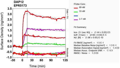
Equilibrium disassociation constant (KD)Learn more about KD Click here to learn more about KD