Anti-CNPase antibody [11-5B]
| Name | Anti-CNPase antibody [11-5B] |
|---|---|
| Supplier | Abcam |
| Catalog | ab6319 |
| Prices | $404.00 |
| Sizes | 100 µg |
| Host | Mouse |
| Clonality | Monoclonal |
| Isotype | IgG1 |
| Clone | 11-5B |
| Applications | IHC-F FC Conjugation DB ELISA ICC/IF IHC-P IHC-F IP WB ICC/IF ICC/IF |
| Species Reactivities | Mouse, Rat, Dog, Human, Monkey, Sheep, Rabbit, Bovine, Pig |
| Antigen | Full length native protein (purified) (Human) |
| Description | Mouse Monoclonal |
| Gene | CNP |
| Conjugate | Unconjugated |
| Supplier Page | Shop |
Product images
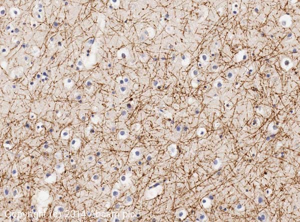 IHC image of CNPase staining in human cerebral cortex formalin fixed paraffin embedded tissue section, performed on a Leica Bond™ system using the standard protocol F. The section was pre-treated using heat mediated antigen retrieval with sodium citrate buffer (pH6, epitope retrieval solution 1) for 20 mins. The section was then incubated with ab6319, 5µg/ml, for 15 mins at room temperature and detected using an HRP conjugated compact polymer system. DAB was used as the chromogen. The section was then counterstained with haematoxylin and mounted with DPX.For other IHC staining systems (automated and non-automated) customers should optimize variable parameters such as antigen retrieval conditions, primary antibody concentration and antibody incubation times.
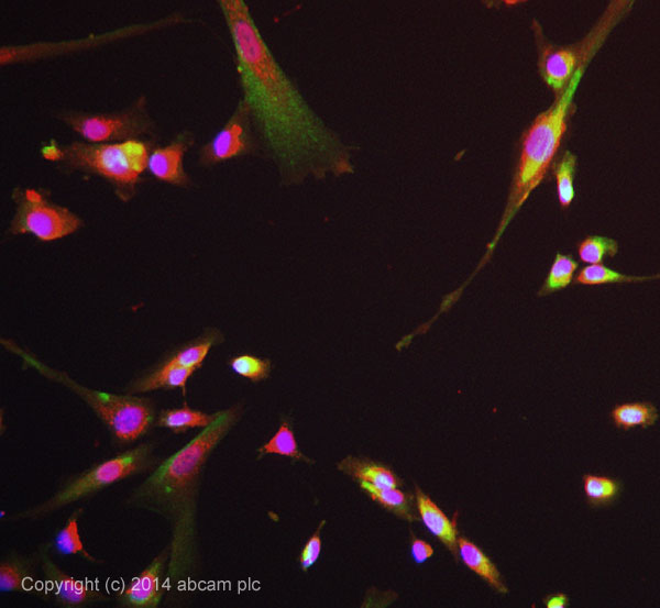 ICC/IF image of ab6319 stained SKNSH cells. The cells were 4% formaldehyde fixed (10 min) and then incubated in 1%BSA / 10% normal goat serum / 0.3M glycine in 0.1% PBS-Tween for 1h to permeabilise the cells and block non-specific protein-protein interactions. The cells were then incubated with the antibody ab6319 at 10µg/ml overnight at +4°C. The secondary antibody (pseudo-colored green) was Alexa Fluor® 488 goat anti- mouse (ab150117) IgG (H+L) preadsorbed, used at a 1/1000 dilution for 1h. Alexa Fluor® 594 WGA was used to label plasma membranes (pseudo-colored red) at a 1/200 dilution for 1h at room temperature. DAPI was used to stain the cell nuclei (pseudo-colored blue) at a concentration of 1.43µM for 1hour at room temperature.
![All lanes : Anti-CNPase antibody [11-5B] (ab6319) at 1/100 dilutionLane 1 : Spinal Cord (Human) Tissue Lysate - adult normal tissue (ab29188)Lane 2 : Brain (Human) Tissue Lysate - adult normal tissue (ab29466)Lane 3 : Spinal Cord (Mouse) Tissue LysateLane 4 : Brain (Mouse) Tissue Lysate Lane 5 : Spinal Cord (Rat) Tissue LysateLane 6 : Brain (Rat) Tissue LysateLysates/proteins at 20 µg per lane.SecondaryGoat polyclonal to Mouse IgG - H&L - Pre-Adsorbed (HRP) at 1/3000 dilutiondeveloped using the ECL techniquePerformed under reducing conditions.](http://www.bioprodhub.com/system/product_images/ab_products/2/sub_2/137_CNPase-Primary-antibodies-ab6319-16.jpg) All lanes : Anti-CNPase antibody [11-5B] (ab6319) at 1/100 dilutionLane 1 : Spinal Cord (Human) Tissue Lysate - adult normal tissue (ab29188)Lane 2 : Brain (Human) Tissue Lysate - adult normal tissue (ab29466)Lane 3 : Spinal Cord (Mouse) Tissue LysateLane 4 : Brain (Mouse) Tissue Lysate Lane 5 : Spinal Cord (Rat) Tissue LysateLane 6 : Brain (Rat) Tissue LysateLysates/proteins at 20 µg per lane.SecondaryGoat polyclonal to Mouse IgG - H&L - Pre-Adsorbed (HRP) at 1/3000 dilutiondeveloped using the ECL techniquePerformed under reducing conditions.
All lanes : Anti-CNPase antibody [11-5B] (ab6319) at 1/100 dilutionLane 1 : Spinal Cord (Human) Tissue Lysate - adult normal tissue (ab29188)Lane 2 : Brain (Human) Tissue Lysate - adult normal tissue (ab29466)Lane 3 : Spinal Cord (Mouse) Tissue LysateLane 4 : Brain (Mouse) Tissue Lysate Lane 5 : Spinal Cord (Rat) Tissue LysateLane 6 : Brain (Rat) Tissue LysateLysates/proteins at 20 µg per lane.SecondaryGoat polyclonal to Mouse IgG - H&L - Pre-Adsorbed (HRP) at 1/3000 dilutiondeveloped using the ECL techniquePerformed under reducing conditions.
 IHC-P image of CNPase staining on rat brain sections using ab6319 (1:1600). heat mediated antigen retrieval on paraffin embedded sections was performed using citric acid. The sections were then blocked with 1% BSA for 10 min at 21°C. The primary antibody was incubated for 16 hours at 21°C. The sections were then incubated in Goat anti-mouse (Biotin) at 1:200. See Abreview
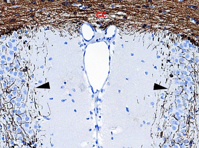
 ab6319 at a 1/2000 dilution staining mouse brain tissue sections by Immunohistochemistry (Frozen sections). The tissue was paraformaldehyde fixed and blocked with 2% BSA. The antibody was incubated with the tissue for 10 hours and then bound antibody was detected using a biotinylated goat anti-mouse antibody.CNPase staining in the striatum is shown in the image.This image is courtesy of an Abreview submitted by Bing Lang on 19 April 2006.See Abreview
 ab6319 at a 1/200 dilution staining rat spinal cord cells by ICC/IF. The cells were ethanol fixed and blocked with 5% serum. The antibody was incubated with the cells for 1 hour and then bound antibody was detected using an Alexa Fluor® 488 conjugated Goat anti-mouse antibody. This image is courtesy of an Abreview submitted by Nancy Nutile-McMenemy.See Abreview
 ab6319 at a 1/200 dilution staining rat spinal cord tissue sections from a 4% PFA transcardially perfused animal by Immunohistochemistry (Frozen sections). The tissue was paraformaldehyde fixed and incubated with the antibody for 18 hours. Bound antibody was detected using an HRP conjugated goat anti-mouse polyclonal antibody. This image is courtesy of an Abreview submitted by Nancy Nutile-McMenemy.See Abreview
![All lanes : Anti-CNPase antibody [11-5B] (ab6319) at 1/500 dilutionLane 1 : 40ug rat brain homegenate (whole tissue lysate) animal #661Lane 2 : 40ug rat brain homegenate (whole tissue lysate) animal #655Lane 3 : 40ug rat brain homegenate (whole tissue lysate) animal #668Lane 4 : 40ug rat brain homegenate (whole tissue lysate) animal #662Lane 5 : 40ug rat brain homegenate (whole tissue lysate) animal #659Lane 6 : 40ug rat brain homegenate (whole tissue lysate) animal #670SecondaryHRP conjugated goat anti-mouse antibodydeveloped using the ECL techniquePerformed under reducing conditions.](http://www.bioprodhub.com/system/product_images/ab_products/2/sub_2/142_ab6319_5.jpg) All lanes : Anti-CNPase antibody [11-5B] (ab6319) at 1/500 dilutionLane 1 : 40ug rat brain homegenate (whole tissue lysate) animal #661Lane 2 : 40ug rat brain homegenate (whole tissue lysate) animal #655Lane 3 : 40ug rat brain homegenate (whole tissue lysate) animal #668Lane 4 : 40ug rat brain homegenate (whole tissue lysate) animal #662Lane 5 : 40ug rat brain homegenate (whole tissue lysate) animal #659Lane 6 : 40ug rat brain homegenate (whole tissue lysate) animal #670SecondaryHRP conjugated goat anti-mouse antibodydeveloped using the ECL techniquePerformed under reducing conditions.
All lanes : Anti-CNPase antibody [11-5B] (ab6319) at 1/500 dilutionLane 1 : 40ug rat brain homegenate (whole tissue lysate) animal #661Lane 2 : 40ug rat brain homegenate (whole tissue lysate) animal #655Lane 3 : 40ug rat brain homegenate (whole tissue lysate) animal #668Lane 4 : 40ug rat brain homegenate (whole tissue lysate) animal #662Lane 5 : 40ug rat brain homegenate (whole tissue lysate) animal #659Lane 6 : 40ug rat brain homegenate (whole tissue lysate) animal #670SecondaryHRP conjugated goat anti-mouse antibodydeveloped using the ECL techniquePerformed under reducing conditions.
![All lanes : Anti-CNPase antibody [11-5B] (ab6319) at 1/750 dilutionLane 1 : Spinal Cord homogenate (whole tissue lysate)Lane 2 : Spinal Cord homogenate (whole tissue lysate)Lane 3 : Spinal Cord homogenate (whole tissue lysate)Lysates/proteins at 2 µg per lane.SecondaryHRP conjugated sheep anti-mouse IgG](http://www.bioprodhub.com/system/product_images/ab_products/2/sub_2/143_ab6319_6.jpg) All lanes : Anti-CNPase antibody [11-5B] (ab6319) at 1/750 dilutionLane 1 : Spinal Cord homogenate (whole tissue lysate)Lane 2 : Spinal Cord homogenate (whole tissue lysate)Lane 3 : Spinal Cord homogenate (whole tissue lysate)Lysates/proteins at 2 µg per lane.SecondaryHRP conjugated sheep anti-mouse IgG
All lanes : Anti-CNPase antibody [11-5B] (ab6319) at 1/750 dilutionLane 1 : Spinal Cord homogenate (whole tissue lysate)Lane 2 : Spinal Cord homogenate (whole tissue lysate)Lane 3 : Spinal Cord homogenate (whole tissue lysate)Lysates/proteins at 2 µg per lane.SecondaryHRP conjugated sheep anti-mouse IgG
 ab6319 staining mouse brain tissue sections by IHC-FoFr. Sections were PFA fixed and permeabilized in 0.1% Triton X-100 prior to blocking in 0.5% TNB for 30 minutes at 25°C. The primary antibody was diluted 1/250 and incubated with the sample for 18 hours at 25°C. An Alexa Fluor® 488 conjugated goat anti-mouse antibody, diluted 1/250, was used as the secondary.Image demonstrates a 2-D depth projection through the superficial cortex.See Abreview
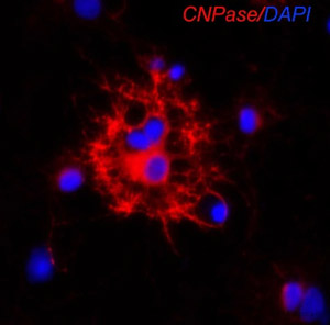 ab6319 staining CNPase in rat oligodendrocytes by Immunocytochemistry/ Immunofluorescence.Cells were fixed in paraformaldehyde, blocked using 5% serum for 10 minutes at 25°C, then incubated with ab6319 at a 1/200 dilution for 2 hours at 25°C. The secondary used was a goat anti-mouse Cy3 conjugated polyclonal at a 1/100 dilution.See Abreview
![Overlay histogram showing SH-SY5Y cells stained with ab6319 (red line). The cells were fixed with 80% methanol (5 min) and then permeabilized with 0.1% PBS-Tween for 20 min. The cells were then incubated in 1x PBS / 10% normal goat serum / 0.3M glycine to block non-specific protein-protein interactions followed by the antibody (ab6319, 2µg/1x106 cells) for 30 min at 22ºC. The secondary antibody used was DyLight® 488 goat anti-mouse IgG (H+L) (ab96879) at 1/500 dilution for 30 min at 22ºC. Isotype control antibody (black line) was mouse IgG1 [ICIGG1] (ab91353, 2µg/1x106 cells) used under the same conditions. Acquisition of >5,000 events was performed. This antibody gave a positive signal in SH-SY5Y cells fixed with 4% paraformaldehyde (10 min)/permeabilized with 0.1% PBS-Tween for 20 min used under the same conditions.](http://www.bioprodhub.com/system/product_images/ab_products/2/sub_2/146_CNPase-Primary-antibodies-ab6319-43.jpg) Overlay histogram showing SH-SY5Y cells stained with ab6319 (red line). The cells were fixed with 80% methanol (5 min) and then permeabilized with 0.1% PBS-Tween for 20 min. The cells were then incubated in 1x PBS / 10% normal goat serum / 0.3M glycine to block non-specific protein-protein interactions followed by the antibody (ab6319, 2µg/1x106 cells) for 30 min at 22ºC. The secondary antibody used was DyLight® 488 goat anti-mouse IgG (H+L) (ab96879) at 1/500 dilution for 30 min at 22ºC. Isotype control antibody (black line) was mouse IgG1 [ICIGG1] (ab91353, 2µg/1x106 cells) used under the same conditions. Acquisition of >5,000 events was performed. This antibody gave a positive signal in SH-SY5Y cells fixed with 4% paraformaldehyde (10 min)/permeabilized with 0.1% PBS-Tween for 20 min used under the same conditions.
Overlay histogram showing SH-SY5Y cells stained with ab6319 (red line). The cells were fixed with 80% methanol (5 min) and then permeabilized with 0.1% PBS-Tween for 20 min. The cells were then incubated in 1x PBS / 10% normal goat serum / 0.3M glycine to block non-specific protein-protein interactions followed by the antibody (ab6319, 2µg/1x106 cells) for 30 min at 22ºC. The secondary antibody used was DyLight® 488 goat anti-mouse IgG (H+L) (ab96879) at 1/500 dilution for 30 min at 22ºC. Isotype control antibody (black line) was mouse IgG1 [ICIGG1] (ab91353, 2µg/1x106 cells) used under the same conditions. Acquisition of >5,000 events was performed. This antibody gave a positive signal in SH-SY5Y cells fixed with 4% paraformaldehyde (10 min)/permeabilized with 0.1% PBS-Tween for 20 min used under the same conditions.
Product References
Fluorescence-activated sorting of fixed nuclei: a general method for studying - Fluorescence-activated sorting of fixed nuclei: a general method for studying
Marion-Poll L, Montalban E, Munier A, Herve D, Girault JA. Eur J Neurosci. 2014 Apr;39(7):1234-44.
Multiple pathogenic proteins implicated in neuronopathic Gaucher disease mice. - Multiple pathogenic proteins implicated in neuronopathic Gaucher disease mice.
Xu YH, Xu K, Sun Y, Liou B, Quinn B, Li RH, Xue L, Zhang W, Setchell KD, Witte D, Grabowski GA. Hum Mol Genet. 2014 Aug 1;23(15):3943-57.
Role of 2',3'-cyclic nucleotide 3'-phosphodiesterase in the renal - Role of 2',3'-cyclic nucleotide 3'-phosphodiesterase in the renal
Jackson EK, Gillespie DG, Mi Z, Cheng D, Bansal R, Janesko-Feldman K, Kochanek PM. Am J Physiol Renal Physiol. 2014 Jul 1;307(1):F14-24. doi:
Cerebrospinal fluid derived from progressive multiple sclerosis patients promotes - Cerebrospinal fluid derived from progressive multiple sclerosis patients promotes
Cristofanilli M, Cymring B, Lu A, Rosenthal H, Sadiq SA. Neuroscience. 2013 Oct 10;250:614-21.
Convection-enhanced delivery of AAV2 in white matter--a novel method for gene - Convection-enhanced delivery of AAV2 in white matter--a novel method for gene
Barua NU, Woolley M, Bienemann AS, Johnson D, Wyatt MJ, Irving C, Lewis O, Castrique E, Gill SS. J Neurosci Methods. 2013 Oct 30;220(1):1-8.
Perinatal inflammation results in decreased oligodendrocyte numbers in adulthood. - Perinatal inflammation results in decreased oligodendrocyte numbers in adulthood.
Graf AE, Haines KM, Pierson CR, Bolon BN, Houston RH, Velten M, Heyob KM, Rogers LK. Life Sci. 2014 Jan 17;94(2):164-71.
Evaluation of novel acyclic nucleoside phosphonates against human and animal - Evaluation of novel acyclic nucleoside phosphonates against human and animal
Coen N, Duraffour S, Naesens L, Krecmerova M, Van den Oord J, Snoeck R, Andrei G. J Virol. 2013 Nov;87(22):12422-32.
Exposure to As, Cd and Pb-mixture impairs myelin and axon development in rat - Exposure to As, Cd and Pb-mixture impairs myelin and axon development in rat
Rai NK, Ashok A, Rai A, Tripathi S, Nagar GK, Mitra K, Bandyopadhyay S. Toxicol Appl Pharmacol. 2013 Dec 1;273(2):242-58. doi:
Oxidative stress and proinflammatory cytokines contribute to demyelination and - Oxidative stress and proinflammatory cytokines contribute to demyelination and
di Penta A, Moreno B, Reix S, Fernandez-Diez B, Villanueva M, Errea O, Escala N, Vandenbroeck K, Comella JX, Villoslada P. PLoS One. 2013;8(2):e54722.
Prolonged myelination in human neocortical evolution. - Prolonged myelination in human neocortical evolution.
Miller DJ, Duka T, Stimpson CD, Schapiro SJ, Baze WB, McArthur MJ, Fobbs AJ, Sousa AM, Sestan N, Wildman DE, Lipovich L, Kuzawa CW, Hof PR, Sherwood CC. Proc Natl Acad Sci U S A. 2012 Oct 9;109(41):16480-5. doi: