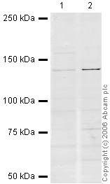
All lanes : Anti-CENPC antibody (ab33034) at 1 µg/mlLane 1 : HeLa (Human epithelial carcinoma cell line) Whole Cell Lysate at 10 µgLane 2 : Jurkat (Human T cell lymphoblast-like cell line) Whole Cell Lysate (ab7899) at 20 µgSecondaryIR Dye 680 Conjugated Goat Anti-Rabbit IgG (H+L) at 1/15000 dilutionPerformed under reducing conditions.

ICC/IF image of ab33034 stained human HeLa cells. The cells were 4% PFA fixed (10 min), permabilised in TBS-T (20 min) and incubated with the antibody (ab33034, 1µg/ml) for 1h at room temperature. 1%BSA / 10% normal goat serum / 0.3M glycine was used to quench autofluorescence and block non-specific protein-protein interactions. The secondary antibody (green) was Alexa Fluor® 488 goat anti-rabbit IgG (H+L) used at a 1/1000 dilution for 1h. Alexa Fluor® 594 WGA was used to label plasma membranes (red). DAPI was used to stain the cell nuclei (blue).

ab33034 (1/200) staining CENPC in HeLa cells (green). Cells were fixed in methanol and counterstained with DAPI in order to highlight the nucleus (red). Please refer to abreview for further experimental details.See Abreview