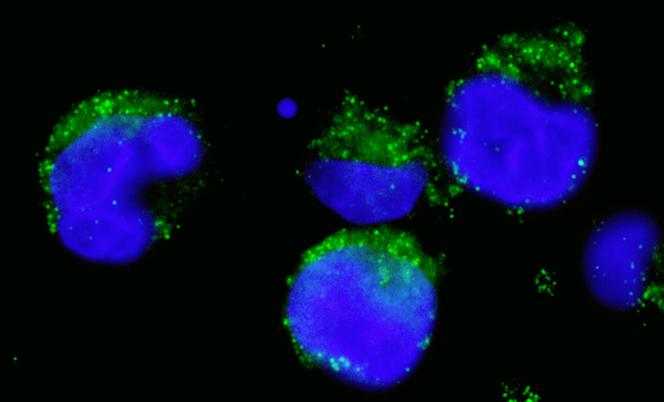
Immunocytochemistry/Immunofluorescence analysis of human K562 cells labelling Bcr with ab86173 at 1/1000 (1µg/ml). An Alexa Fluor® 488-conjugated goat anti-rabbit IgG (1/1000) was used as the secondary antibody.
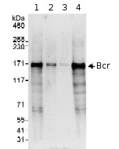
All lanes : Anti-Bcr antibody (ab86173) at 0.1 µg/mlLane 1 : HeLa whole cell lysate at 50 µgLane 2 : HeLa whole cell lysate at 15 µgLane 3 : HeLa whole cell lysate at 5 µgLane 4 : 293T whole cell lysate at 50 µgdeveloped using the ECL technique
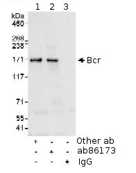
Detection of Human Bcr by Immunoprecipitation, using ab86173 at 10µg/mg lysate. Image shows immunoprecipitated Bcr detected with post IP WB, loading 20% of IP and using HeLa whole cell lysate at 1mg, with an antibody recognising an upstream epitope in lane 1, ab86173 at 1 µg/ml in lane 2 and control IgG in lane 3. Detection: Chemiluminescence with an exposure time of 10 seconds.
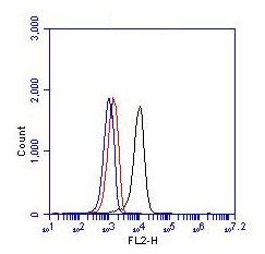
Flow Cytometric Detection of Bcr. 1 x 10⁶ K562 cells were fixed, permeabilized, and stained with ab86173 at 0.25 µg in a 150 µl reaction. Image shows Isotype anti-KLH control (red), no antibody (blue) and ab86173 (black).
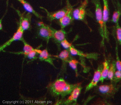
ICC/IF image of ab86173 stained HepG2 cells. The cells were 4% formaldehyde (10 min) and then incubated in 1%BSA / 10% normal goat serum / 0.3M glycine in 0.1% PBS-Tween for 1h to permeabilise the cells and block non-specific protein-protein interactions. The cells were then incubated with the antibody (ab86173, 5µg/ml) overnight at +4°C. The secondary antibody (green) was ab96899 Dylight 488 goat anti-rabbit IgG (H+L) used at a 1/250 dilution for 1h. Alexa Fluor® 594 WGA was used to label plasma membranes (red) at a 1/200 dilution for 1h. DAPI was used to stain the cell nuclei (blue) at a concentration of 1.43µM.