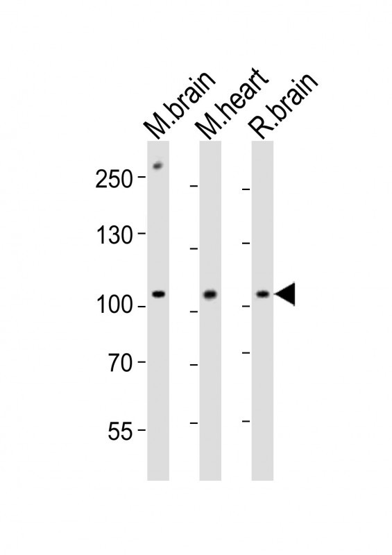
All lanes : Anti-Ephb1 Antibody (Center) at 1:1000 dilution Lane 1: mouse brain lysates Lane 2: mouse heart lysates Lane 3: rat brain lysates Lysates/proteins at 20 µg per lane. Secondary Goat Anti-Rabbit IgG, (H+L), Peroxidase conjugated at 1/10000 dilution Predicted band size : 110 kDa Blocking/Dilution buffer: 5% NFDM/TBST.

Western blot analysis of lysates from A431, mouse NIH/3T3 cell line, mouse brain, rat brain tissue lysate(from left to right), using Ephb1 Antibody (Center)(Cat. #AP20994a). AP20994a was diluted at 1:1000 at each lane. A goat anti-rabbit IgG H&L(HRP) at 1:10000 dilution was used as the secondary antibody. Lysates at 20ug per lane.