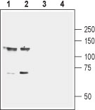
Western blot analysis of mouse (lanes 1 and 3) and rat (lanes 2 and 4) brain membranes: 1-2. Anti-EphB1 (extracellular) antibody (#AG1432), (1:800). 3-4. Anti-EphB1 (extracellular) antibody, preincubated with the control peptide antigen.

Expression of EphB1 in rat PC12 cells Immunocytochemical staining of live intact rat PC12 pheochromocytoma cells. A. Extracellular staining of cells with Anti-EphB1 (extracellular) antibody (#AG1432), (1:100), followed by goat anti-rabbit-AlexaFluor- 594 (red). B. Live image of the cells. C. Merge of the two images.
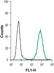
Indirect flow cytometry analysis of live intact human THP-1 monocytic leukemia cell line: ___ Cells + goat anti-rabbit-AlexaFluor-488 secondary antibody.___ Cells + Anti-EphB1 (extracellular) antibody (#AG1432), (1:15) + goat-anti-rabbit-Alexa-488.