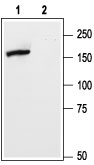
Western blot analysis of rat brain membranes: 1. Anti-mGluR5 (extracellular) antibody (#AG1262), (1:500). 2. Anti-mGluR5 (extracellular) antibody, preincubated with the control peptide antigen.
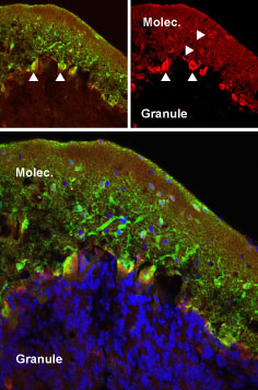
Expression of mGluR5 in rat cerebellum and hippocampus Immunohistochemical staining of perfusion-fixed frozen rat cerebellum sections using Anti-mGluR5 (extracellular) antibody (#AG1262), (1:50). mGluR5 (red) was detected in cerebellar Purkinje cells (vertical arrows) and in the molecular layer (horizontal arrows). Staining with mouse anti-parvalbumin (green) revealed co-localization in Purkinje but not in the molecular layer. Little staining of mGluR5 was detected in the granule layer. DAPI counterstain is used to visualize nuclei of all cells (blue).
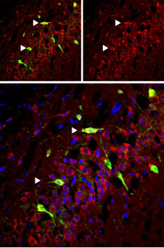
Expression of mGluR5 in rat cerebellum and hippocampus Immunohistochemical staining of perfusion-fixed frozen rat hippocampus sections using Anti-mGluR5 (extracellular) antibody (#AG1262), (1:50). mGluR5 (red) was detected in CA3 cells (arrows). Staining with mouse anti-parvalbumin (green) revealed co-localization in pyramidal layer. DAPI counterstain was used to visualize nuclei of all cells (blue)
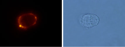
Expression of mGluR5 in rat GH3 pituitary cells Immunocytochemical staining of live intact rat GH3 pituitary cells using Anti-mGluR5 (extracellular) antibody (#AG1262), (1:100), followed by goat-anti-rabbit-AlexaFluor-555 secondary antibody.