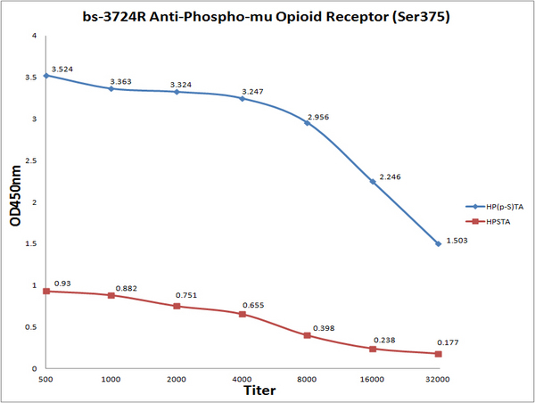
Antigen: Phospho Mu Opioid Receptor (blue line), 0.2ug/100ul; Mu Opioid Receptor(red line), 0.2ug/100ul\\nPrimary: Antiserum, 1:500, 1:1000, 1:2000, 1:4000, 1:8000, 1:16000, 1:32000; \\nSecondary: HRP conjugated Goat Anti-Rabbit IgG(bs-0295G-HRP) at 1: 5000; \\nTMB staining; \\nRead the data in MicroplateReader by 450nm. \\n

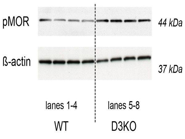
Image kindly provided by Dr. Stefen Clemens of East Carolina University. pMOR protein expression levels in the spinal cords of WT and D3KO mice (top lanes), and their respective ß-actin protein expression (bottom panels). Rabbit Anti-mu Opioid Receptor (Ser375) Polyclonal Antibody was used at a dilution of 1:1000.

