Anti-YWHAZ / 14-3-3 Zeta Antibody (aa65-93)
| Name | Anti-YWHAZ / 14-3-3 Zeta Antibody (aa65-93) |
|---|---|
| Supplier | LifeSpan Bioscience |
| Catalog | LS-C160705 |
| Prices | $295.00 |
| Sizes | 400 µl |
| Host | Rabbit |
| Clonality | Polyclonal |
| Applications | IHC-P WB |
| Species Reactivities | Human, Mouse, Rat |
| Purity/Format | Protein A purified |
| Blocking Peptide | YWHAZ / 14-3-3 Zeta Antibody Blocking Peptide |
| Description | Rabbit Polyclonal |
| Gene | YWHAZ |
| Conjugate | Unconjugated |
| Supplier Page | Shop |
Product images
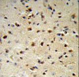 YWHAZ Antibody IHC of formalin-fixed and paraffin-embedded mouse brain followed by peroxidase-conjugated secondary antibody and DAB staining.
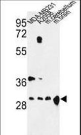 Western blot of YWHAZ Antibody in MDA-MB231, A2058 cell line and mouse cerebellum, brain tissue lysates (35 ug/lane). YWHAZ (arrow) was detected using the purified antibody.
 YWHAZ Antibody flow cytometry of MDA-MB231 cells (right histogram) compared to a negative control cell (left histogram). FITC-conjugated goat-anti-rabbit secondary antibodies were used for the analysis.