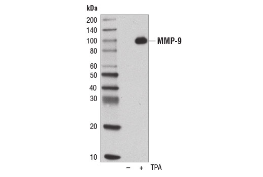
Western blot analysis of concentrated, serum-free cultured medium from U-2 OS cells, untreated (-) or treated with TPA #4174 (200 nM, 48 hr; +), using MMP-9 (D6O3H) XP ® Rabbit mAb.

Immunohistochemical analysis of paraffin-embedded human breast carcinoma using MMP-9 (D6O3H) XP ® Rabbit mAb.

Immunohistochemical analysis of paraffin-embedded human lung carcinoma using MMP-9 (D6O3H) XP ® Rabbit mAb.

Immunohistochemical analysis of paraffin-embedded U-2 OS cell pellets, untreated (left) or treated with TPA #4174 (right), using MMP-9 (D6O3H) XP ® Rabbit mAb.

Immunohistochemical analysis of paraffin-embedded human colon carcinoma using MMP-9 (D6O3H) XP ® Rabbit mAb in the presence of control peptide (left) or antigen-specific peptide (right).

Confocal immunofluorescent analysis of serum starved U-2 OS cells, untreated (left) or treated with TPA #4174 (200 nM, 24 hr; right), using MMP-9 (D6O3H) XP ® Rabbit mAb (green). Blue pseudocolor = DRAQ5 ® #4084 (fluorescent DNA dye).

Flow cytometric analysis of serum starved U-2 OS cells, untreated (blue) or treated with TPA (200nM, 24 hours) using MMP-9 (D6O3H) XP Rabbit mAb (green). Anti-rabbit IgG (H+L), F(ab')2 Fragment (PE Conjugate) was used as a secondary antibody.