Phospho-IκBα (Ser32/36) (5A5) Mouse mAb
| Name | Phospho-IκBα (Ser32/36) (5A5) Mouse mAb |
|---|---|
| Supplier | Cell Signaling Technology |
| Catalog | 9246 |
| Prices | $287.00, $678.00 |
| Sizes | 100 µl (10 western blots), 300 µl (30 western blots) |
| Host | Mouse |
| Clonality | Monoclonal |
| Isotype | IgG1 |
| Clone | 5A5 |
| Applications | WB |
| Species Reactivities | Human, Mouse, Rat, Monkey, Bovine, Dog, Pig, Guinea Pig |
| Antigen | Monoclonal antibody is produced by immunizing animals with a synthetic phosphopeptide corresponding to residues surrounding Ser32/36 of human IκBα. |
| Description | Mouse Monoclonal |
| Gene | NFKBIA |
| Supplier Page | Shop |
Product images
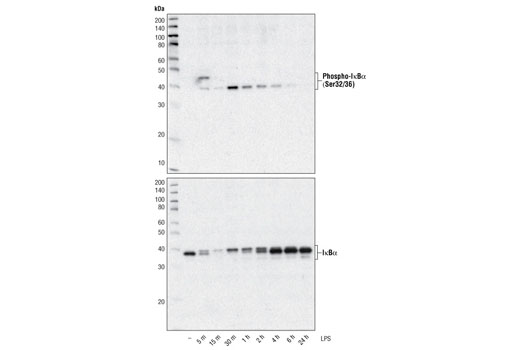 Western blot analysis of extracts from THP-1 cells, differentiated with TPA (#9905, 80 nM for 24 h) and treated with 1 μg/ml LPS for the indicated times, using Phospho-IκBα (Ser32/36) (5A5) Mouse mAb (upper) and IκBα (L35A5) Mouse mAb (Amino-terminal Antigen) #4814 (lower).
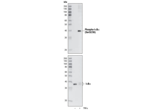 Western blot analysis of extracts from NIH/3T3 cells, untreated orTNF-α-treated (#2169, 20 ng/ml) for 5 minutes, using Phospho-IκBα (Ser32/36) (5A5) Mouse mAb (upper) or IκBα (L35A5) Mouse mAb (Amino-terminal Antigen) #4814 (lower).