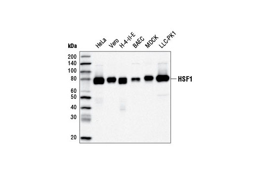
Western blot analysis of extracts from various cell lines using HSF1 (D3L8I) Rabbit mAb.

Immunoprecipitation of HSF1 from HeLa cell extracts using Rabbit (DA1E) mAb IgG XP ® Isotype Control #3900 (lane 2), HSF1 Antibody #4356 (lane 3) or HSF1 (D8L8I) Rabbit mAb (lane 4). Lane 1 is 10% input. Western blot analysis was performed using HSF1 (D8L8I) Rabbit mAb.

Immunohistochemical analysis of paraffin-embedded human lung carcinoma using HSF1 (D3L8I) Rabbit mAb.

Immunohistochemical analysis of paraffin-embedded human colon carcinoma using HSF1 (D3L8I) Rabbit mAb in the presence of control peptide (left) or antigen-specific peptide (right).

Immunohistochemical analysis of paraffin-embedded human prostate carcinoma using HSF1 (D3L8I) Rabbit mAb.

Immunohistochemical analysis of paraffin-embedded mouse spleen, HSF1 wild type (left), HSF1 knock out (middle) or HSF2 knock out (right), using HSF1 (D3L8I) Rabbit mAb. Tissues courtesy of Dr. Nahid Mivechi, Georgia Health Sciences University, Atlanta, Georgia.

Confocal immunofluorescent analysis of HeLa cells, untreated or heat-treated (43.5ºC, 1 hr without recovery), using HSF1 (D3L8I) Rabbit mAb (green). Actin filaments were labeled with DyLight® 554 Phalloidin #13054 (red). HSF1 is redistributed to nuclear stress granules following heat shock (insets).

Flow cytometric analysis of HeLa cells using HSF1 (D3L8I) Rabbit mAb (blue) compared to Rabbit (DA1E) mAb IgG XP ® Isotype Control #3900 (red). Anti-rabbit IgG (H+L), F(ab') 2 Fragment (Alexa Fluor ® 488 Conjugate) #4412 was used as a secondary antibody.

Chromatin immunoprecipitations were performed with cross-linked chromatin from 4 x 10 6 HeLa cells heat shocked for 1h and either 10 µl of HSF1 (D3L8I) Rabbit mAb or 2 µl of Normal Rabbit IgG #2729 using SimpleChIP ® Enzymatic Chromatin IP Kit (Magnetic Beads) #9003. The enriched DNA was quantified by real-time PCR using SimpleChIP ® Human HSPA6 Promoter Primers #5551, human HSP70 intron 1 primers, and SimpleChIP ® Human α Satellite Repeat Primers #4486. The amount of immunoprecipitated DNA in each sample is represented as signal relative to the total amount of input chromatin, which is equivalent to one.