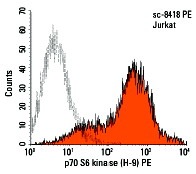
p70 S6 kinase α (H-9) PE: sc-8418 PE. Intracellular FCM analysis of fixed and permeabilized Jurkat cells. Black line histogram represents the isotype control, normal mouse IgG
1: sc-2866.
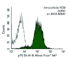
p70 S6 kinase (H-9) Alexa Fluor 647: sc-8418 AF647. Intracellular FCM analysis of fixed and permeabilized Jurkat cells. Black line histogram represents the isotype control, normal mouse IgG
1: sc-24636.
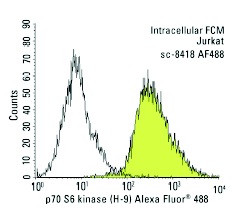
p70 S6 kinase (H-9) Alexa Fluor 488: sc-8418 AF488. Intracellular FCM analysis of fixed and permeabilized Jurkat cells. Black line histogram represents the isotype control, normal mouse IgG
1: sc-3890.
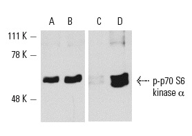
Western blot analysis of p-p70 S6 kinase α activation in whole cell lysates from serum-starved (A,C) and serum-starved then serum-treated (B,D) NIH/3T3 cells. Antibodies tested include p-p70 S6 kinase α (H-9): sc-8418 (A,B) and p-p70 S6 kinase α (A-6): sc-8416 (C,D).
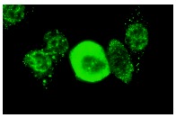
p70 S6 kinase α (H-9): sc-8418. Immunofluorescence staining of methanol-fixed NIH/3T3 cells showing cytoplasmic and nuclear localization.
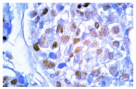
p70 S6 kinase α (H-9): sc-8418. Immunoperoxidase staining of formalin-fixed, paraffin-embedded human breast carcinoma showing nuclear localization.

p70 S6 kinase α (H-9): sc-8418. Western blot analysis of p70 S6 kinase α expression in non-transfected 293T: sc-117752 (A), mouse p70 S6 kinase α transfected 293T: sc-125770 (B) and NIH/3T3 (C) whole cell lysates.
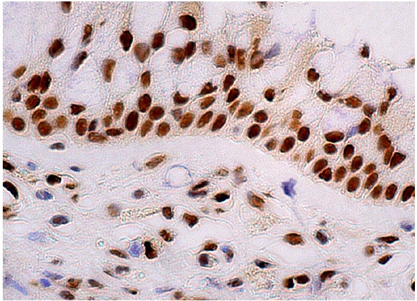
p70 S6 kinase α (H-9): sc-8418. Immunoperoxidase staining of formalin fixed, paraffin-embedded human nasopharynx tissue showing nuclear staining of respiratory epithelial cells.