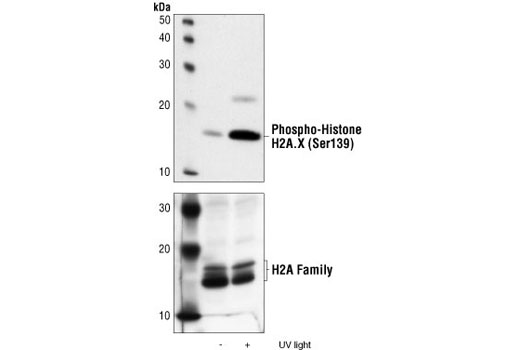
Western blot analysis of extracts from 293 cells, untreated or UV-treated, using Phospho-Histone H2A.X (Ser139) Antibody (upper) or Histone H2A Antibody #2572 (lower).

Confocal microscopic images of HeLa cells, UV treated (A) and untreated (B), showing nuclear stain with Phospho-Histone H2A.X (Ser139) Antibody (red) and Phospho-SAPK/JNK (Thr183/Tyr185) (G9) Mouse mAb #9255 (green).

Flow cytometric analysis of HeLa cells, untreated (blue) and UV-treated (green), using Phospho-Histone H2A.X (Ser139) Antibody compared with a nonspecific negative control antibody (red).


