Anti-ARFGAP1 Antibody (clone 1H6)
| Name | Anti-ARFGAP1 Antibody (clone 1H6) |
|---|---|
| Supplier | LifeSpan Bioscience |
| Catalog | LS-C172746 |
| Prices | $325.00 |
| Sizes | 100 µl |
| Host | Mouse |
| Clonality | Monoclonal |
| Isotype | IgG1 |
| Clone | 1H6 |
| Applications | ICC/IF WB FC |
| Species Reactivities | Human, Monkey |
| Antigen | ARFGAP1 antibody was raised against full length human recombinant protein of human ARFGAP1 (NP_783202) produced in HEK293T cell. |
| Purity/Format | Protein A/G purified |
| Description | Mouse Monoclonal |
| Gene | ARFGAP1 |
| Conjugate | Unconjugated |
| Supplier Page | Shop |
Product images
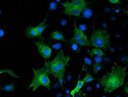 Anti-ARFGAP1 mouse monoclonal antibody immunofluorescent staining of COS7 cells transiently transfected by pCMV6-ENTRY ARFGAP1.
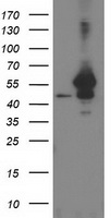 HEK293T cells were transfected with the pCMV6-ENTRY control (Left lane) or pCMV6-ENTRY ARFGAP1 (Right lane) cDNA for 48 hrs and lysed. Equivalent amounts of cell lysates (5 ug per lane) were separated by SDS-PAGE and immunoblotted with anti-ARFGAP1.
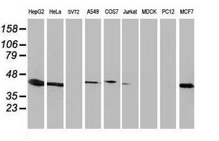 Western blot of extracts (35 ug) from 9 different cell lines by using anti-ARFGAP1 monoclonal antibody.
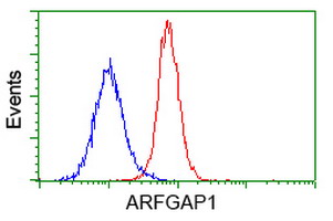
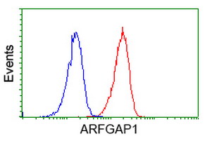 Flow cytometry of Jurkat cells, using anti-ARFGAP1 antibody (Red), compared to a nonspecific negative control antibody (Blue).
 Flow cytometry of HeLa cells, using anti-ARFGAP1 antibody (Red), compared to a nonspecific negative control antibody (Blue).