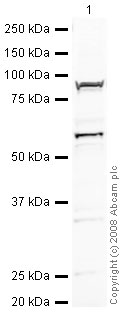
Anti-YY1 antibody - Nuclear Loading Control (ab43058) at 2 µg/ml + Jurkat (Human T cell lymphoblast-like cell line) Nuclear Lysate (ab14844) at 20 µgSecondaryIRDye 680 Conjugated Goat Anti-Rabbit IgG (H+L) at 1/10000 dilutionPerformed under reducing conditions.
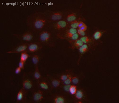
ICC/IF image of ab43058 stained human HEK 293 cells. The cells were 4% PFA fixed (10 min), permabilised in 0.1% PBS-Tween (20 min) and incubated with the antibody (ab43058, 1µg/ml) for 1h at room temperature. 1%BSA / 10% normal goat serum / 0.3M glycine was used to block non-specific protein-protein interactions. The secondary antibody (green) was Alexa Fluor® 488 goat anti-rabbit IgG (H+L) used at a 1/1000 dilution for 1h. Alexa Fluor® 594 WGA was used to label plasma membranes (red). DAPI was used to stain the cell nuclei (blue). This antibody also gave a positive IF result in HeLa, HepG2 and MCF7 cells.
.jpg)
IHC image of YY1 staining in human breast carcinoma FFPE section, performed on a BondTM system using the standard protocol F. The section was pre-treated using heat mediated antigen retrieval with sodium citrate buffer (pH6, epitope retrieval solution 1) for 20 mins. The section was then incubated with ab43058, 5µg/ml, for 8 mins at room temperature and detected using an HRP conjugated compact polymer system. DAB was used as the chromogen. The section was then counterstained with haematoxylin and mounted with DPX.