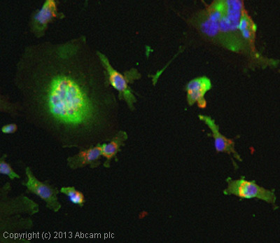
ICC/IF image of ab9959 stained MDA-MB-231 cells. The cells were 4% formaldehyde fixed (10 min) and then incubated in 1%BSA / 10% normal goat serum / 0.3M glycine in 0.1% PBS-Tween for 1h to permeabilise the cells and block non-specific protein-protein interactions. The cells were then incubated with the antibody ab9959 at 1µg/ml overnight at +4°C. The secondary antibody (green) was DyLight® 488 goat anti- rabbit (ab96899) IgG (H+L) used at a 1/250 dilution for 1h. Alexa Fluor® 594 WGA was used to label plasma membranes (red) at a 1/200 dilution for 1h. DAPI was used to stain the cell nuclei (blue) at a concentration of 1.43µM.
![Standard curve for TRAIL (Analyte: ab78818); dilution range 1pg/ml to 1µg/ml using Capture Antibody Mouse monoclonal [55B709.3] to TRAIL (ab12124) at 0.2µg/ml and Detector Antibody Rabbit polyclonal to TRAIL (ab9959) at 0.1µg/ml.](http://www.bioprodhub.com/system/product_images/ab_products/2/sub_5/13820_TRAIL-Primary-antibodies-ab9959-2.jpg)
Standard curve for TRAIL (Analyte: ab78818); dilution range 1pg/ml to 1µg/ml using Capture Antibody Mouse monoclonal [55B709.3] to TRAIL (ab12124) at 0.2µg/ml and Detector Antibody Rabbit polyclonal to TRAIL (ab9959) at 0.1µg/ml.

ab9959 staining TRAIL in human metastatic carcinoma of lymph nodes from breast tissue by Immunohistochemistry (Formalin/PFA fixed paraffin-embedded sections). Tissue underwent heat mediated antigen retrieval in sodium citrate buffer (pH 6.0). The primary antibody was used at 0.25 ug/ml and incubated with sample at 4°C overnight. A HRP-labeled polymer detection system was used with a DAB chromogen.

ab9959 staining TRAIL in human metastatic carcinoma of lymph nodes from breast tissue by Immunohistochemistry (Formalin/PFA fixed paraffin-embedded sections). Tissue underwent heat mediated antigen retrieval in sodium citrate buffer (pH 6.0). The primary antibody was used at 0.25 ug/ml and incubated with sample at 4°C overnight. A HRP-labeled polymer detection system was used with a DAB chromogen.