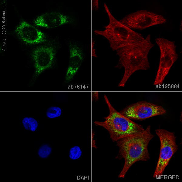
ab76147 staining TRAF3 in Malme-3 cells. The cells were fixed with 4% formaldehyde (10min), permeabilized with 0.1% Triton X-100 for 5 minutes and then blocked with 1% BSA/10% normal goat serum/0.3M glycine in 0.1%PBS-Tween for 1h. The cells were then incubated overnight at +4°C with ab76147 at 1/2000 dilution and ab195884, Rat monoclonal to Tubulin (Alexa Fluor® A647) at 1µg/ml. This was followed by an incubation at room temperature for 1h with ab150081, Goat polyclonal Secondary Antibody to Rabbit IgG (Alexa Fluor® A488) , at 1µg/ml (shown in green). Nuclear DNA was labelled with DAPI (shown in blue).Image was taken with a confocal microscope (Leica-Microsystems, TCS SP8).
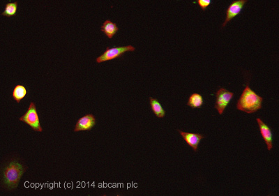
ICC/IF image of ab76147 stained MALME-3M cells. The cells were 4% formaldehyde fixed (10 min) and then incubated in 1%BSA / 10% normal goat serum / 0.3M glycine in 0.1% PBS-Tween for 1h to permeabilise the cells and block non-specific protein-protein interactions. The cells were then incubated with the antibody ab76147 at 1/100 dilution overnight at +4°C. The secondary antibody (pseudo-colored green) was Alexa Fluor® 488 goat anti- rabbit (ab150081) IgG (H+L) preadsorbed, used at a 1/1000 dilution for 1h. Alexa Fluor® 594 WGA was used to label plasma membranes (pseudo-colored red) at a 1/200 dilution for 1h at room temperature. DAPI was used to stain the cell nuclei (pseudo-colored blue) at a concentration of 1.43µM for 1hour at room temperature.
![Anti-TRAF3 antibody [EP1730Y] (ab76147) at 1/500 dilution + Molt-4 cell lysate at 10 µg](http://www.bioprodhub.com/system/product_images/ab_products/2/sub_5/13713_ab76147_1.jpg)
Anti-TRAF3 antibody [EP1730Y] (ab76147) at 1/500 dilution + Molt-4 cell lysate at 10 µg

ab76147, at 1/50 dilution, staining TRAF3 in paraffin-embedded human tonsil tissue by immunohistochemistry.
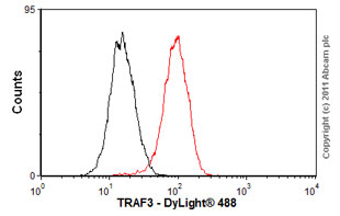
Overlay histogram showing Jurkat cells stained with ab76147 (red line). The cells were fixed with 80% methanol (5 min) and then permeabilized with 0.1% PBS-Tween for 20 min. The cells were then incubated in 1x PBS / 10% normal goat serum / 0.3M glycine to block non-specific protein-protein interactions followed by the antibody (ab76147, 1/100 dilution) for 30 min at 22ºC. The secondary antibody used was DyLight® 488 goat anti-rabbit IgG (H+L) (ab96899) at 1/500 dilution for 30 min at 22ºC. Isotype control antibody (black line) was rabbit IgG (monoclonal) (1µg/1x106 cells) used under the same conditions. Acquisition of >5,000 events was performed.