![All lanes : Anti-TBP like protein TLP [EPR16048] antibody (ab191391) at 1/20000 dilutionLane 1 : HeLa cell lysateLane 2 : HepG2 cell lysateLane 3 : 293T cell lysateLysates/proteins at 20 µg per lane.SecondaryGoat Anti-Rabbit IgG, (H+L),Peroxidase conjugated at 1/1000 dilution](http://www.bioprodhub.com/system/product_images/ab_products/2/sub_5/8935_ab191391-228653-ab191391WB.jpg)
All lanes : Anti-TBP like protein TLP [EPR16048] antibody (ab191391) at 1/20000 dilutionLane 1 : HeLa cell lysateLane 2 : HepG2 cell lysateLane 3 : 293T cell lysateLysates/proteins at 20 µg per lane.SecondaryGoat Anti-Rabbit IgG, (H+L),Peroxidase conjugated at 1/1000 dilution
![All lanes : Anti-TBP like protein TLP [EPR16048] antibody (ab191391) at 1/10000 dilutionLane 1 : Mouse testis lysateLane 2 : Rat testis lysateLysates/proteins at 10 µg per lane.SecondaryGoat Anti-Rabbit IgG, (H+L),Peroxidase conjugated at 1/1000 dilution](http://www.bioprodhub.com/system/product_images/ab_products/2/sub_5/8936_ab191391-228652-ab191391WBb.jpg)
All lanes : Anti-TBP like protein TLP [EPR16048] antibody (ab191391) at 1/10000 dilutionLane 1 : Mouse testis lysateLane 2 : Rat testis lysateLysates/proteins at 10 µg per lane.SecondaryGoat Anti-Rabbit IgG, (H+L),Peroxidase conjugated at 1/1000 dilution
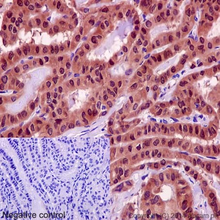
Immunohistochemical analysis of paraffin-embedded Human thyroid carcinoma tissue labeling TBP like protein TLP with ab191391 at 1/100 dilution, followed by HRP Polymer for Rabbit/Mouse IgG. Counter stained with Hematoxylin.Negative control: PBS instead of primary antibody.
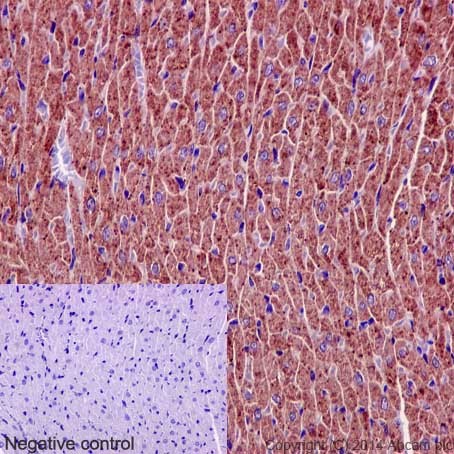
Immunohistochemical analysis of paraffin-embedded Human cardiac muscle tissue labeling TBP like protein TLP with ab191391 at 1/100 dilution, followed by HRP Polymer for Rabbit/Mouse IgG. Counter stained with Hematoxylin.Negative control: PBS instead of primary antibody.
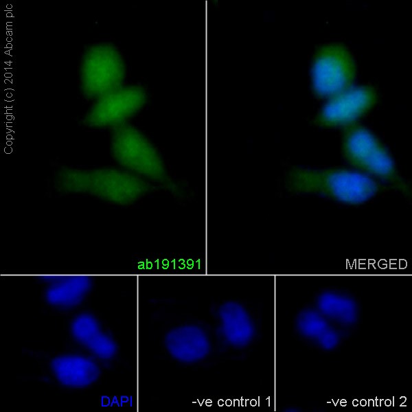
Immunofluorescent analysis of 4% paraformaldehyde-fixed, 0.1% tritonX-100 permeabilized HeLa cells labeling TBP like protein TLP with ab191391 at 1/250 dilution, followed by Goat anti rabbit IgG (Alexa Fluor® 488) secondary antibody (ab150077) at 1/200 dilution (green). Nuclear counter stain is DAPI (blue).The negative controls are as follows;-ve control 1 and -ve control 2 - ab191391 at 1/250 dilution followed by Goat anti mouse IgG (Alexa Fluor®594) secondary antibody at 1/400 dilution.
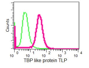
Flow cytometric analysis of 2% paraformaldehyde-fixed 293T cells labeling TBP like protein TLP with ab191391 at 1/110 dilution (pink), compared to a Rabbit monoclonal IgG isotype control (green). Goat anti rabbit IgG (FITC) secondary antibody was used at 1/150 dilution.
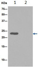
Western blot analysis of TBP like protein TLP in 293T cell lysate immunoprecipitated with ab191391 at 1/30 dilution (Lane 1). Lane 2: PBS instead of 293T cell lysate.Secondary antibody: Anti-Rabbit IgG (HRP), specific to the non-reduced form of IgG at 1/1500 dilution.Blocking/dilution buffer: 5% NFDM/TBST.