Anti-SPHK1 antibody
| Name | Anti-SPHK1 antibody |
|---|---|
| Supplier | Abcam |
| Catalog | ab16491 |
| Prices | $400.00 |
| Sizes | 100 µg |
| Host | Rabbit |
| Clonality | Polyclonal |
| Isotype | IgG |
| Applications | IHC-P WB ICC/IF ICC/IF |
| Species Reactivities | Mouse, Rat, Human |
| Antigen | Synthetic peptide conjugated to KLH derived from within residues 350 to the C-terminus of Human SPHK1 |
| Blocking Peptide | Human SPHK1 peptide |
| Description | Rabbit Polyclonal |
| Gene | SPHK1 |
| Conjugate | Unconjugated |
| Supplier Page | Shop |
Product images
 All lanes : Anti-SPHK1 antibody (ab16491) at 1 µg/mlLane 1 : Tagged Recombinant SPHK1Lane 2 : Tagged Recombinant SPHK1 with Human SPHK1 peptide (ab16634) at 1 µg/mlLysates/proteins at 0.1 µg per lane.SecondaryAlexa Fluor Goat polyclonal to Rabbit IgG (700) at 1/10000 dilution
 All lanes : Anti-SPHK1 antibody (ab16491) at 1 µg/mlLane 1 : Human Skeletal muscle lysateLane 2 : Human Skeletal muscle lysate with Human SPHK1 peptide (ab16634) at 1 µg/mlLysates/proteins at 20 µg/ml per lane.SecondaryAlexa Fluor Goat polyclonal to Rabbit IgG (700) at 1/10000 dilution
 ab16491 staining human smooth muscle, mouse lung, and rat heart tissue sections by IHC-P. Sections were formaldehyde fixed and subjected to heat mediated antigen retrieval in citrate buffer (pH 6) prior to blocking in sequential peroxidase and protein block (prediluted) for 20 minutes at 20°C. The primary antibody was diluted 1/100 and incubated with the sample for 45 minutes at 20°C. A HRP-conjugated goat anti-rabbit antibody was used as the secondary.See Abreview
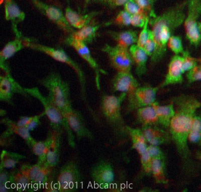 ICC/IF image of ab16491 stained HepG2 cells. The cells were 4% PFA fixed (10 min) and then incubated in 1%BSA / 10% normal goat serum / 0.3M glycine in 0.1% PBS-Tween for 1h to permeabilise the cells and block non-specific protein-protein interactions. The cells were then incubated with the antibody (ab16491, 5µg/ml) overnight at +4°C. The secondary antibody (green) was ab96899 Dylight 488 goat anti-rabbit IgG (H+L) used at a 1/250 dilution for 1h. Alexa Fluor® 594 WGA was used to label plasma membranes (red) at a 1/200 dilution for 1h. DAPI was used to stain the cell nuclei (blue) at a concentration of 1.43µM.
Product References
Inhibition of insulin-like growth factor-binding protein-3 signaling through - Inhibition of insulin-like growth factor-binding protein-3 signaling through
Martin JL, de Silva HC, Lin MZ, Scott CD, Baxter RC. Mol Cancer Ther. 2014 Feb;13(2):316-28.
Intracellular S1P generation is essential for S1P-induced motility of human lung - Intracellular S1P generation is essential for S1P-induced motility of human lung
Berdyshev EV, Gorshkova I, Usatyuk P, Kalari S, Zhao Y, Pyne NJ, Pyne S, Sabbadini RA, Garcia JG, Natarajan V. PLoS One. 2011 Jan 31;6(1):e16571.
S1P lyase regulates DNA damage responses through a novel sphingolipid feedback - S1P lyase regulates DNA damage responses through a novel sphingolipid feedback
Kumar A, Oskouian B, Fyrst H, Zhang M, Paris F, Saba JD. Cell Death Dis. 2011 Feb 10;2:e119.