![All lanes : Anti-SERPINB1 antibody [EPR13305(B)] (ab181084) at 1/1000 dilutionLane 1 : Human fetal lung tissue lysateLane 2 : MCF7 whole cell lysateLane 3 : HeLa whole cell lysateLysates/proteins at 20 µg per lane.](http://www.bioprodhub.com/system/product_images/ab_products/2/sub_4/28655_ab181084-214773-ab181084WB.jpg)
All lanes : Anti-SERPINB1 antibody [EPR13305(B)] (ab181084) at 1/1000 dilutionLane 1 : Human fetal lung tissue lysateLane 2 : MCF7 whole cell lysateLane 3 : HeLa whole cell lysateLysates/proteins at 20 µg per lane.
![Anti-SERPINB1 antibody [EPR13305(B)] (ab181084) at 1/1000 dilution + HepG2 whole cell lysate at 20 µg](http://www.bioprodhub.com/system/product_images/ab_products/2/sub_4/28656_ab181084-214774-ab181084WB2.jpg)
Anti-SERPINB1 antibody [EPR13305(B)] (ab181084) at 1/1000 dilution + HepG2 whole cell lysate at 20 µg

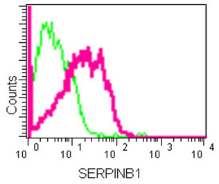
Flow cytometric analysis of HepG2 cells fixed with 2% paraformaldehyde labeling SERPINB1 with ab181084 at 1/70 dilution (red), compared to a rabbit IgG negative control (green).

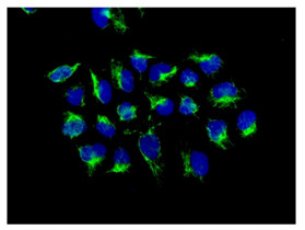
Immunofluorescent analysis of HeLa cells labeling SERPINB1 with ab181084 at a 1/100 dilution. Primary incubation was followed by secondary antibody labeling with Alexa Fluor 488-conjugated goat anti-rabbit lgG (green). DAPI was used to stain the cell nuclear (blue).

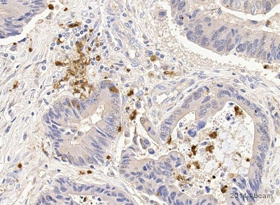
ab181084 staining SERPINB1 in human colorectal cancer tissue sections by Immunohistochemistry (IHC-P - paraformaldehyde-fixed, paraffin-embedded sections). Tissue was fixed with formaldehyde, permeabilized with 0.2% Triton X-100 in PBS and blocked with 5% milk for 30 minutes at room temperature; antigen retrieval was by heat mediation in Tris/HCl pH 9.0. Samples were incubated with primary antibody (1/500) for 1 hour. An undiluted HRP-conjugated Goat anti-rabbit IgG polyclonal was used as the secondary antibody.See Abreview
![All lanes : Anti-SERPINB1 antibody [EPR13305(B)] (ab181084) at 1/1000 dilutionLane 1 : Human fetal lung tissue lysateLane 2 : MCF7 whole cell lysateLane 3 : HeLa whole cell lysateLysates/proteins at 20 µg per lane.](http://www.bioprodhub.com/system/product_images/ab_products/2/sub_4/28655_ab181084-214773-ab181084WB.jpg)
![Anti-SERPINB1 antibody [EPR13305(B)] (ab181084) at 1/1000 dilution + HepG2 whole cell lysate at 20 µg](http://www.bioprodhub.com/system/product_images/ab_products/2/sub_4/28656_ab181084-214774-ab181084WB2.jpg)


