Anti-S100 beta antibody [EP1576Y]
| Name | Anti-S100 beta antibody [EP1576Y] |
|---|---|
| Supplier | Abcam |
| Catalog | ab52642 |
| Prices | $403.00 |
| Sizes | 100 µl |
| Host | Rabbit |
| Clonality | Monoclonal |
| Isotype | IgG |
| Clone | EP1576Y |
| Applications | ICC/IF ICC/IF WB IP IHC-P |
| Species Reactivities | Mouse, Rat, Goat, Human, Zebrafish, Monkey |
| Antigen | Synthetic peptide (the amino acid sequence is considered to be commercially sensitive) corresponding to Human S100 beta aa 50 to the C-terminus (C terminal) |
| Description | Rabbit Monoclonal |
| Gene | S100B |
| Conjugate | Unconjugated |
| Supplier Page | Shop |
Product images
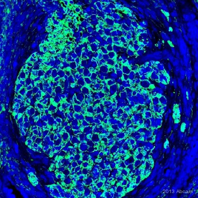
 Immunofluorescent analysis of 4% paraformaldehyde-fixed, 0.1% Triton X-100 A-375 (Human malignant melanoma cell line) cells labeling S100 beta with purified ab52642 at 1/100 dilution, followed by Goat anti rabbit IgG (Alexa Fluor® 488) ab150077 secondary antibody at 1/500 dilution (green). The nuclear counter stain is DAPI (blue). The negative control is as follows;ab52642 at 1/100 dilution followed by ab150120 (AlexaFluor®594 Goat anti-Mouse secondary) at 1/500 dilution.
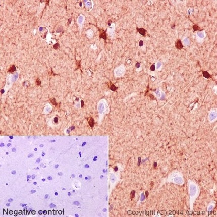 Immunohistochemical analysis of paraffin embedded Human cerebral cortex tissue labeling S100 beta with purified ab52642 at 1/1000 dilution. Secondary antibody was Goat Anti-Rabbit IgG H&L (HRP) (ab97051) at 1/500 dilution. Counter stain: Hematoxylin.Negative control: Using PBS instead of primary antibody.
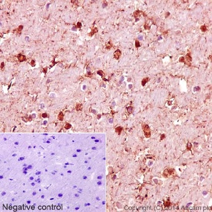 Immunohistochemical analysis of paraffin embedded Rat cerebral cortex tissue labeling S100 beta with purified ab52642 at 1/1000 dilution. Secondary antibody was Goat Anti-Rabbit IgG H&L (HRP) (ab97051) at 1/500 dilution. Counter stain: Hematoxylin.Negative control: Using PBS instead of primary antibody.
 Immunohistochemical analysis of paraffin embedded Mouse cerebral cortex tissue labeling S100 beta with purified ab52642 at 1/1000 dilution. Secondary antibody was Goat Anti-Rabbit IgG H&L (HRP) (ab97051) at 1/500 dilution. Counter stain: Hematoxylin.Negative control: Using PBS instead of primary antibody.
![Anti-S100 beta antibody [EP1576Y] (ab52642) at 1/5000 dilution (purified) + B16F0 (Mouse melanoma cell line) at 10 µgSecondaryGoat Anti-Rabbit IgG, (H+L), Peroxidase conjugated at 1/1000 dilution](http://www.bioprodhub.com/system/product_images/ab_products/2/sub_4/26117_ab52642-238773-ab52642.jpg) Anti-S100 beta antibody [EP1576Y] (ab52642) at 1/5000 dilution (purified) + B16F0 (Mouse melanoma cell line) at 10 µgSecondaryGoat Anti-Rabbit IgG, (H+L), Peroxidase conjugated at 1/1000 dilution
Anti-S100 beta antibody [EP1576Y] (ab52642) at 1/5000 dilution (purified) + B16F0 (Mouse melanoma cell line) at 10 µgSecondaryGoat Anti-Rabbit IgG, (H+L), Peroxidase conjugated at 1/1000 dilution
![All lanes : Anti-S100 beta antibody [EP1576Y] (ab52642) at 1/10000 dilution (purified)Lane 1 : Mouse spinal cordLane 2 : Rat brainLysates/proteins at 20 µg per lane.SecondaryGoat Anti-Rabbit IgG, (H+L), Peroxidase conjugated at 1/1000 dilution](http://www.bioprodhub.com/system/product_images/ab_products/2/sub_4/26118_ab52642-238772-ab52642.jpg) All lanes : Anti-S100 beta antibody [EP1576Y] (ab52642) at 1/10000 dilution (purified)Lane 1 : Mouse spinal cordLane 2 : Rat brainLysates/proteins at 20 µg per lane.SecondaryGoat Anti-Rabbit IgG, (H+L), Peroxidase conjugated at 1/1000 dilution
All lanes : Anti-S100 beta antibody [EP1576Y] (ab52642) at 1/10000 dilution (purified)Lane 1 : Mouse spinal cordLane 2 : Rat brainLysates/proteins at 20 µg per lane.SecondaryGoat Anti-Rabbit IgG, (H+L), Peroxidase conjugated at 1/1000 dilution
![Anti-S100 beta antibody [EP1576Y] (ab52642) at 1/5000 dilution (purified) + A-375 (Human malignant melanoma cell line) at 10 µgSecondaryGoat Anti-Rabbit IgG, (H+L), Peroxidase conjugated at 1/1000 dilution](http://www.bioprodhub.com/system/product_images/ab_products/2/sub_4/26119_ab52642-238771-ab52642.jpg) Anti-S100 beta antibody [EP1576Y] (ab52642) at 1/5000 dilution (purified) + A-375 (Human malignant melanoma cell line) at 10 µgSecondaryGoat Anti-Rabbit IgG, (H+L), Peroxidase conjugated at 1/1000 dilution
Anti-S100 beta antibody [EP1576Y] (ab52642) at 1/5000 dilution (purified) + A-375 (Human malignant melanoma cell line) at 10 µgSecondaryGoat Anti-Rabbit IgG, (H+L), Peroxidase conjugated at 1/1000 dilution
 S100 beta was immunoprecipitated from Human fetal brain with purified ab52642 at 1/50 dilution. Western blot was performed from the immunoprecipitate using ab52642 and Goat Anti-Rabbit IgG, (H+L), Peroxidase conjugated was used as secondary antibody at 1/1000 dilution.Blocking and dilution buffer and concentration: 5% NFDM/TBST.
![Anti-S100 beta antibody [EP1576Y] (ab52642) at 1/1000 dilution (unpurified) + A375 cell lysate at 10 µgSecondaryGoat anti-Rabbit HRP labeled at 1/2000 dilution](http://www.bioprodhub.com/system/product_images/ab_products/2/sub_4/26121_ab52642_1.bmp) Anti-S100 beta antibody [EP1576Y] (ab52642) at 1/1000 dilution (unpurified) + A375 cell lysate at 10 µgSecondaryGoat anti-Rabbit HRP labeled at 1/2000 dilution
Anti-S100 beta antibody [EP1576Y] (ab52642) at 1/1000 dilution (unpurified) + A375 cell lysate at 10 µgSecondaryGoat anti-Rabbit HRP labeled at 1/2000 dilution
 Unpurified ab52642 staining S100 beta in Human spiral ganglion tissue sections by Immunohistochemistry (IHC-P - paraformaldehyde-fixed, paraffin-embedded sections). Tissue was fixed with paraformaldehyde and blocked with 1% BSA for 30 minutes at room temperature; antigen retrieval was by heat mediation in citrate buffer, pH6.0. Samples were incubated with primary antibody (1/200 in PBS-T + 1% BSA) for 12 hours. An Alexa Fluor® 488-conjugated Donkey anti-rabbit IgG polyclonal (1/500) was used as the secondary antibody.See Abreview
 Unpurified ab52642 at 1/500 staining human melanoma tissue sections by IHC-P. The tissue was formladehyde fixed and a heat mediated antigen retrieval step was performed, before being blocked and incubated with the antibody for 1 hour. An HRP conjugated goat anti-rabbit antibody was used as the secondary.See Abreview
 Immunohistochemical analysis of embryonic mouse brain tissue, staining S100 beta with unpurified ab52642.
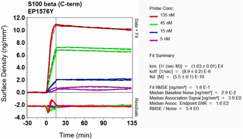 Equilibrium disassociation constant (KD)Learn more about KD Click here to learn more about KD
Product References
Distribution and development of peripheral glial cells in the human fetal - Distribution and development of peripheral glial cells in the human fetal
Locher H, de Groot JC, van Iperen L, Huisman MA, Frijns JH, Chuva de Sousa Lopes SM. PLoS One. 2014 Jan 31;9(1):e88066.
S100A9 knockout decreases the memory impairment and neuropathology in crossbreed - S100A9 knockout decreases the memory impairment and neuropathology in crossbreed
Kim HJ, Chang KA, Ha TY, Kim J, Ha S, Shin KY, Moon C, Nacken W, Kim HS, Suh YH. PLoS One. 2014 Feb 25;9(2):e88924.
RAGE regulates immune cell infiltration and angiogenesis in choroidal - RAGE regulates immune cell infiltration and angiogenesis in choroidal
Chen M, Glenn JV, Dasari S, McVicar C, Ward M, Colhoun L, Quinn M, Bierhaus A, Xu H, Stitt AW. PLoS One. 2014 Feb 26;9(2):e89548.
Sirtuin modulators control reactive gliosis in an in vitro model of Alzheimer's - Sirtuin modulators control reactive gliosis in an in vitro model of Alzheimer's
Scuderi C, Stecca C, Bronzuoli MR, Rotili D, Valente S, Mai A, Steardo L. Front Pharmacol. 2014 May 13;5:89.
Distinct spatial distribution of microglia and macrophages following mesenchymal - Distinct spatial distribution of microglia and macrophages following mesenchymal
Le Blon D, Hoornaert C, Daans J, Santermans E, Hens N, Goossens H, Berneman Z, Ponsaerts P. Immunol Cell Biol. 2014 Sep;92(8):650-8.
Regulation of S100B in white adipose tissue by obesity in mice. - Regulation of S100B in white adipose tissue by obesity in mice.
Buckman LB, Anderson-Baucum EK, Hasty AH, Ellacott KLj. Adipocyte. 2014 Jul 1;3(3):215-20.
Biochemical assessment of precuneus and posterior cingulate gyrus in the context - Biochemical assessment of precuneus and posterior cingulate gyrus in the context
Maarouf CL, Kokjohn TA, Walker DG, Whiteside CM, Kalback WM, Whetzel A, Sue LI, Serrano G, Jacobson SA, Sabbagh MN, Reiman EM, Beach TG, Roher AE. PLoS One. 2014 Aug 28;9(8):e105784.
Sustained down-regulation of beta-dystroglycan and associated dysfunctions of - Sustained down-regulation of beta-dystroglycan and associated dysfunctions of
Gondo A, Shinotsuka T, Morita A, Abe Y, Yasui M, Nuriya M. J Biol Chem. 2014 Oct 31;289(44):30279-88.
Lack of evidence for vesicular glutamate transporter expression in mouse - Lack of evidence for vesicular glutamate transporter expression in mouse
Li D, Herault K, Silm K, Evrard A, Wojcik S, Oheim M, Herzog E, Ropert N. J Neurosci. 2013 Mar 6;33(10):4434-55.
3-D imaging and illustration of mouse intestinal neurovascular complex. - 3-D imaging and illustration of mouse intestinal neurovascular complex.
Fu YY, Peng SJ, Lin HY, Pasricha PJ, Tang SC. Am J Physiol Gastrointest Liver Physiol. 2013 Jan 1;304(1):G1-11. doi: