Immunoprecipitation of Rad51 protein from crude extract of HeLa cells by ab179897.ab179897 (20 µg) was incubated with 20 µg of HeLa cell extract, and precipitated with 20 μg of protein A-beads. The sample was dissociated from the precipitate by heating in SDS-sample buffer and analyzed by western blotting with anti-Rad51 antiserum (chicken, ab63802) at 1/1000 dilution. As secondary antibody, anti-chicken IgG antibody (rabbit) was used at 1/10000 dilution.
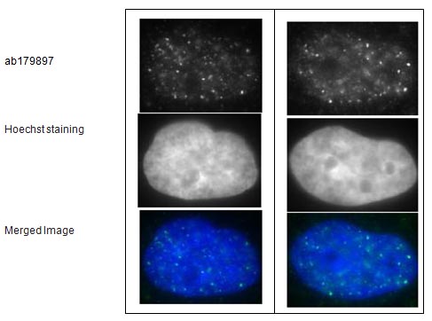
Immunofluorescent detection of Ra51 foci formation after X-ray irradiation in human fibroblast cell line, GM0637.Cells were irradiated by X-rays at 2 Gy, grown for 1 hr, fixed with 4% paraformaldehyde in 1x PBS for 10 min, washed 3 times with PBS for 3 min, permealized by treatment with 0.5% Triton for 5 min, washed 3 times with PBS for 3 min, incubated with ab179897 for 30 min at 37°C, washed 3 times with PBS for 3 min, incubated with secondary antibody for 30 min at 37°C, washed 3 times with PBS for 3 min, stained with Hoechst for 1 min and mounted. ab179897 was used at 1/10000 dilution (left panels) and 1/1000 dilution (right panels). As the secondary antibody, anti-rabbit Alexa 488 was used at 1/10000 dilution.The pictures were by courtesy of Prof. S. Tashiro and Dr. K. Kono at Hiroshima University.
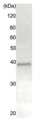
 All lanes : Anti-Rad51 antibody - ChIP Grade (ab179897) at 1/1000 dilutionLane 1 : HeLa crude extract from cells grown in the absence of adriamycin (DNA damaging agent)Lane 2 : HeLa crude extract from cells grown in the presence of adriamycin (DNA damaging agent)Lane 3 : NIH 3T3 crude extract