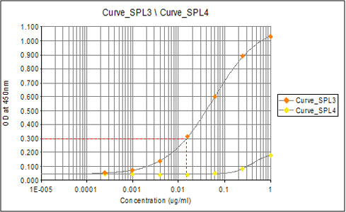
Elisa using ab31769 at varying antibody concentrations. Curve_SPL3 indicates binding to the Rad51 (phospho T309) peptide. Curve_SPL4 indicates <2% crossreactivity with the unmodified Rad51 control peptide.
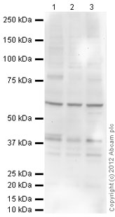
All lanes : Anti-Rad51 (phospho T309) antibody (ab31769) at 1 µg/mlLane 1 : Jurkat (Human T cell lymphoblast-like cell line) Whole Cell Lysate Lane 2 : Ramos (Human Burkitt's lymphoma cell line) Whole Cell Lysate Lane 3 : Y79 (Human retinoblastoma cell line) Whole Cell Lysate Lysates/proteins at 10 µg per lane.SecondaryGoat Anti-Rabbit IgG H&L (HRP) preadsorbed (ab97080) at 1/10000 dilutiondeveloped using the ECL techniquePerformed under reducing conditions.Observed band size : 39 kDa (why is the actual band size different from the predicted?)Additional bands at : 34 kDa,60 kDa. We are unsure as to the identity of these extra bands.Exposure time : 20 minutes
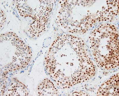
IHC image of Rad51 (phospho T309) staining in human testis formalin fixed paraffin embedded tissue section, performed on a Leica BondTM system using the standard protocol F. The section was pre-treated using heat mediated antigen retrieval with sodium citrate buffer (pH6, epitope retrieval solution 1) for 20 mins. The section was then incubated with ab31769, 1µg/ml, for 15 mins at room temperature and detected using an HRP conjugated compact polymer system. DAB was used as the chromogen. The section was then counterstained with haematoxylin and mounted with DPX. For other IHC staining systems (automated and non-automated) customers should optimize variable parameters such as antigen retrieval conditions, primary antibody concentration and antibody incubation times.