Anti-Histone H2B antibody - ChIP Grade
| Name | Anti-Histone H2B antibody - ChIP Grade |
|---|---|
| Supplier | Abcam |
| Catalog | ab1790 |
| Prices | $403.00 |
| Sizes | 100 µg |
| Host | Rabbit |
| Clonality | Polyclonal |
| Isotype | IgG |
| Applications | WB IHC-P ChIP ICC/IF ICC/IF IP |
| Species Reactivities | Mouse, Rat, Chicken, Bovine, Human, Yeast, Xenopus, Arabidopsis thaliana, C. elegans, Zebrafish |
| Antigen | Synthetic peptide conjugated to KLH derived from within residues 100 to the C-terminus of Human Histone H2B |
| Description | Rabbit Polyclonal |
| Gene | HIST2H2BD |
| Conjugate | Unconjugated |
| Supplier Page | Shop |
Product images
 developed using the ECL techniquePerformed under reducing conditions.
![Histone H2B - ChIP Grade was immunoprecipitated using 0.5mg HeLa whole cell extract, 5µg of Rabbit polyclonal to and 50µl of protein G magnetic beads (+). No antibody was added to the control (-).The antibody was incubated under agitation with Protein G beads for 10min, HeLa whole cell extract lysate diluted in RIPA buffer was added to each sample and incubated for a further 10min under agitation.Proteins were eluted by addition of 40µl SDS loading buffer and incubated for 10min at 70°C; 10µl of each sample was separated on a SDS PAGE gel, transferred to a nitrocellulose membrane, blocked with 5% BSA and probed with ab1790.Secondary: Mouse monoclonal [SB62a] Secondary Antibody to Rabbit IgG light chain (HRP) (ab99697).Band: 14kDa; Histone H2B - ChIP Grade](http://www.bioprodhub.com/system/product_images/ab_products/2/sub_3/1450_ab1790-220131-EpiIP01ab17903m.jpg) Histone H2B - ChIP Grade was immunoprecipitated using 0.5mg HeLa whole cell extract, 5µg of Rabbit polyclonal to and 50µl of protein G magnetic beads (+). No antibody was added to the control (-).The antibody was incubated under agitation with Protein G beads for 10min, HeLa whole cell extract lysate diluted in RIPA buffer was added to each sample and incubated for a further 10min under agitation.Proteins were eluted by addition of 40µl SDS loading buffer and incubated for 10min at 70°C; 10µl of each sample was separated on a SDS PAGE gel, transferred to a nitrocellulose membrane, blocked with 5% BSA and probed with ab1790.Secondary: Mouse monoclonal [SB62a] Secondary Antibody to Rabbit IgG light chain (HRP) (ab99697).Band: 14kDa; Histone H2B - ChIP Grade
Histone H2B - ChIP Grade was immunoprecipitated using 0.5mg HeLa whole cell extract, 5µg of Rabbit polyclonal to and 50µl of protein G magnetic beads (+). No antibody was added to the control (-).The antibody was incubated under agitation with Protein G beads for 10min, HeLa whole cell extract lysate diluted in RIPA buffer was added to each sample and incubated for a further 10min under agitation.Proteins were eluted by addition of 40µl SDS loading buffer and incubated for 10min at 70°C; 10µl of each sample was separated on a SDS PAGE gel, transferred to a nitrocellulose membrane, blocked with 5% BSA and probed with ab1790.Secondary: Mouse monoclonal [SB62a] Secondary Antibody to Rabbit IgG light chain (HRP) (ab99697).Band: 14kDa; Histone H2B - ChIP Grade
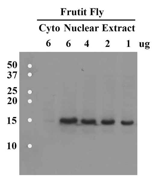
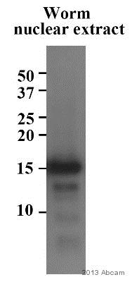 Anti-Histone H2B antibody - ChIP Grade (ab1790) at 1/500 dilution + Caenorhabditis elegans embryo tissue lysate - nuclear at 30 µgSecondaryHRP-conjugated Goat anti-rabbit IgG polyclonal at 1/1000 dilutiondeveloped using the ECL techniquePerformed under reducing conditions.
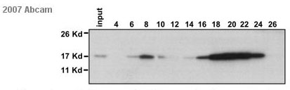
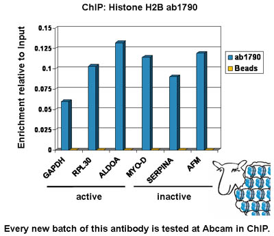 Chromatin was prepared from U2OS cells according to the Abcam X-ChIP protocol. Cells were fixed with formaldehyde for 10min. The ChIP was performed with 25µg of chromatin, 2µg of ab1790 (blue), and 20µl of Protein A/G sepharose beads. No antibody was added to the beads control (yellow). The immunoprecipitated DNA was quantified by real time PCR (Taqman approach for active and inactive loci, Sybr green approach for heterochromatic loci). Primers and probes are located in the first kb of the transcribed region.
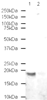 Lane 1 : Anti-Histone H2B antibody - ChIP Grade (ab1790) at 0.1 µg/ml (Sample: Calf thymus histone prep, 20 ug)Lane 2 : Anti-Histone H2B antibody - ChIP Grade (ab1790) at 0.1 µg/mlLane 1 : As aboveLane 2 : Human Histone H2B peptide (ab16101) at 1 µg/mlSecondaryAlexa fluor Goat polyclonal anti-Rabbit IgG at 1/10000 dilutionPerformed under reducing conditions.
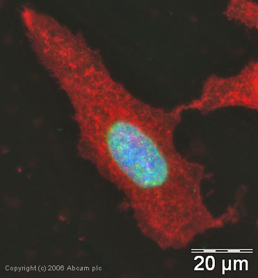 ICC/IF image of ab1790 stained human HeLa cells. The cells were PFA fixed (3.7% PFA, 5 min) and incubated with the antibody (ab1790, 1µg/ml) for 1h at room temperature. The secondary antibody (green) was Alexa Fluor® 488 goat anti-rabbit IgG (H+L) used at a 1/1000 dilution for 1h. Image-iTTM FX Signal Enhancer was used as the primary blocking agent, 5% BSA (in TBS-T) was used for all other blocking steps. DAPI was used to stain the cell nuclei (blue). Alexa Fluor® 594 WGA was used to label plasma membranes (red).
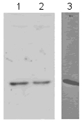 All lanes : Anti-Histone H2B antibody - ChIP Grade (ab1790)Lane 1 : Hela Histone prepLane 2 : Hela whole cell lysateLane 3 : S. cerevisiae whole cell lysatePerformed under reducing conditions.
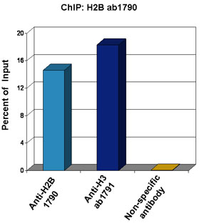 Chromatin from Xenopus laevis oocytes was prepared according to the Abcam X-ChIP protocol. Oocytes were fixed with formaldehyde for 10 min. The ChIP was performed with 25 µg of chromatin, 3 µg of ab1790 (anti-H2B, light blue) and 3 µg of ab1791 (anti-H3, dark blue), and 20 µl of Protein A/G sepharose beads. A non-specific antibody was used as a control (yellow). The immunoprecipitated DNA was quantified by real time PCR (Taqman approach).
 HeLa cells were fixed in 100% methanol for 6 minutes at -20°C. The cells were washed 3 times in PBS then incubated with ab1790 (0.5µg/ml) for 1 hour at room temperature. The panel of images shows the cells stained with ab1790 (green) and counterstained with DAPI (blue). 100x magnification.
 developed using the ECL techniquePerformed under reducing conditions.
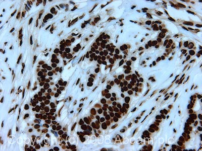 IHC image of Histone H2B staining in human breast carcinoma FFPE section, performed on a BondTM system using the standard protocol F. The section was pre-treated using heat mediated antigen retrieval with sodium citrate buffer (pH6, epitope retrieval solution 1) for 20 mins. The section was then incubated with ab1790, 1µg/ml, for 8 mins at room temperature and detected using an HRP conjugated compact polymer system. DAB was used as the chromogen. The section was then counterstained with haematoxylin and mounted with DPX.
Product References
Human cytomegalovirus major immediate early 1 protein targets host chromosomes by - Human cytomegalovirus major immediate early 1 protein targets host chromosomes by
Mucke K, Paulus C, Bernhardt K, Gerrer K, Schon K, Fink A, Sauer EM, Asbach-Nitzsche A, Harwardt T, Kieninger B, Kremer W, Kalbitzer HR, Nevels M. J Virol. 2014 Jan;88(2):1228-48.
Smc5/6-mediated regulation of replication progression contributes to chromosome - Smc5/6-mediated regulation of replication progression contributes to chromosome
Gallego-Paez LM, Tanaka H, Bando M, Takahashi M, Nozaki N, Nakato R, Shirahige K, Hirota T. Mol Biol Cell. 2014 Jan;25(2):302-17.
SUPT6H controls estrogen receptor activity and cellular differentiation by - SUPT6H controls estrogen receptor activity and cellular differentiation by
Bedi U, Scheel AH, Hennion M, Begus-Nahrmann Y, Ruschoff J, Johnsen SA. Oncogene. 2015 Jan 22;34(4):465-73.
TFPI1 mediates resistance to doxorubicin in breast cancer cells by inducing a - TFPI1 mediates resistance to doxorubicin in breast cancer cells by inducing a
Davies GF, Berg A, Postnikoff SD, Wilson HL, Arnason TG, Kusalik A, Harkness TA. PLoS One. 2014 Jan 28;9(1):e84611.
NuMA promotes homologous recombination repair by regulating the accumulation of - NuMA promotes homologous recombination repair by regulating the accumulation of
Vidi PA, Liu J, Salles D, Jayaraman S, Dorfman G, Gray M, Abad P, Moghe PV, Irudayaraj JM, Wiesmuller L, Lelievre SA. Nucleic Acids Res. 2014 Jun;42(10):6365-79.
Identification of histone 3 variant 2 interacting factors. - Identification of histone 3 variant 2 interacting factors.
Latreille D, Bluy L, Benkirane M, Kiernan RE. Nucleic Acids Res. 2014 Apr;42(6):3542-50.
ATR pathway inhibition is synthetically lethal in cancer cells with ERCC1 - ATR pathway inhibition is synthetically lethal in cancer cells with ERCC1
Mohni KN, Kavanaugh GM, Cortez D. Cancer Res. 2014 May 15;74(10):2835-45.
SETD2 is required for DNA double-strand break repair and activation of the - SETD2 is required for DNA double-strand break repair and activation of the
Carvalho S, Vitor AC, Sridhara SC, Martins FB, Raposo AC, Desterro JM, Ferreira J, de Almeida SF. Elife. 2014 May 6;3:e02482.
Nucleosome assembly is required for nuclear pore complex assembly in mouse - Nucleosome assembly is required for nuclear pore complex assembly in mouse
Inoue A, Zhang Y. Nat Struct Mol Biol. 2014 Jul;21(7):609-16.
.