Anti-Histone H2B antibody [mAbcam 52484] - ChIP Grade
| Name | Anti-Histone H2B antibody [mAbcam 52484] - ChIP Grade |
|---|---|
| Supplier | Abcam |
| Catalog | ab52484 |
| Prices | $403.00 |
| Sizes | 100 µg |
| Host | Mouse |
| Clonality | Monoclonal |
| Isotype | IgG1 |
| Clone | mAbcam 52484 |
| Applications | IP FC WB ICC/IF ICC/IF ChIP IHC-P IHC-F |
| Species Reactivities | Mouse, Rat, Chicken, Human, Drosophila, Zebrafish, Bovine, Xenopus, C. elegans, Orangutan |
| Antigen | Synthetic peptide corresponding to Human Histone H2B aa 100 to the C-terminus (C terminal) conjugated to Keyhole Limpet Haemocyanin (KLH) |
| Description | Mouse Monoclonal |
| Gene | HIST2H2BD |
| Conjugate | Unconjugated |
| Supplier Page | Shop |
Product images
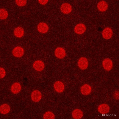 ab52484 staining Histone H2B in Fruit fly (Drosophila melanogaster) embryo cells by ICC/IF (Immunocytochemistry/immunofluorescence). Embryos were washed in 1x PBS + 0.1% Trition, cells were fixed with heat, and blocked with 10% BSA for 12 hours at 4°C. Samples were incubated with primary antibody (1/1000) for 24 hours. An Alexa Fluor®594-conjugated Rabbit anti-mouse IgG polyclonal (1/5000) was used as the secondary antibody.See Abreview
![All lanes : Anti-Histone H2B antibody [mAbcam 52484] - ChIP Grade (ab52484) at 5 µg/mlLane 1 : Histone H1 recombinant protein.Lane 2 : Histone H2A recombinant protein.Lane 3 : Histone H2B recombinant protein.Lane 4 : Histone H3 recombinant protein.Lane 5 : Histone H4 recombinant protein.Lysates/proteins at 0.1 µg per lane.SecondaryRabbit polyclonal to Mouse IgG - H&L (HRP) at 1/3000 dilutionPerformed under reducing conditions.](http://www.bioprodhub.com/system/product_images/ab_products/2/sub_3/1435_ab52484_1.jpg) All lanes : Anti-Histone H2B antibody [mAbcam 52484] - ChIP Grade (ab52484) at 5 µg/mlLane 1 : Histone H1 recombinant protein.Lane 2 : Histone H2A recombinant protein.Lane 3 : Histone H2B recombinant protein.Lane 4 : Histone H3 recombinant protein.Lane 5 : Histone H4 recombinant protein.Lysates/proteins at 0.1 µg per lane.SecondaryRabbit polyclonal to Mouse IgG - H&L (HRP) at 1/3000 dilutionPerformed under reducing conditions.
All lanes : Anti-Histone H2B antibody [mAbcam 52484] - ChIP Grade (ab52484) at 5 µg/mlLane 1 : Histone H1 recombinant protein.Lane 2 : Histone H2A recombinant protein.Lane 3 : Histone H2B recombinant protein.Lane 4 : Histone H3 recombinant protein.Lane 5 : Histone H4 recombinant protein.Lysates/proteins at 0.1 µg per lane.SecondaryRabbit polyclonal to Mouse IgG - H&L (HRP) at 1/3000 dilutionPerformed under reducing conditions.
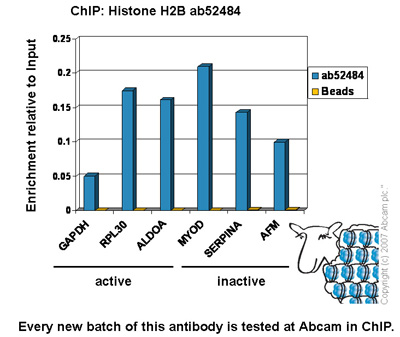 Chromatin was prepared from HeLa cells according to the Abcam X-ChIP protocol. Cells were fixed with formaldehyde for 10min. The ChIP was performed with 25µg of chromatin, 5µg of ab52484 (blue), and 20µl of Protein A/G sepharose beads. No antibody was added to the beads control (yellow). The immunoprecipitated DNA was quantified by real time PCR (Taqman approach). Primers and probes are located in the first kb of the transcribed region.
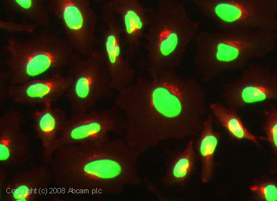 ICC/IF image of ab52484 stained human HeLa cells. The cells were 4% PFA fixed (10 min), permabilised in 0.1% PBS-Tween (20 min) and incubated with the antibody (ab52484, 1µg/ml) for 1h at room temperature. 1%BSA / 10% normal goat serum / 0.3M glycine was used to block non-specific protein-protein interactions. The secondary antibody (green) was Alexa Fluor® 488 goat anti-mouse IgG (H+L) used at a 1/1000 dilution for 1h. Alexa Fluor® 594 WGA was used to label plasma membranes (red). DAPI was used to stain the cell nuclei (blue).
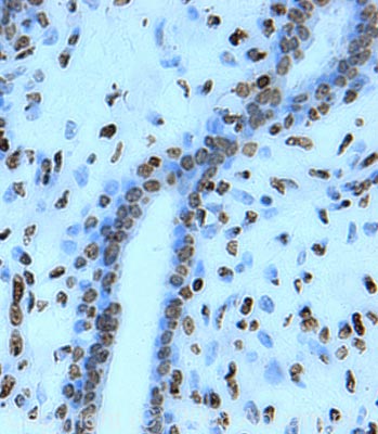 IHC image of Histone H2B staining in human breast carcinoma FFPE section, performed on a BondTM system using the standard protocol F. The section was pre-treated using heat mediated antigen retrieval with sodium citrate buffer (pH6, epitope retrieval solution 1) for 20 mins. The section was then incubated with ab52484, 1µg/ml, for 15 mins at room temperature and detected using an HRP conjugated compact polymer system. DAB was used as the chromogen. The section was then counterstained with haematoxylin and mounted with DPX.
![All lanes : Anti-Histone H2B antibody [mAbcam 52484] - ChIP Grade (ab52484) at 5 µg/mlLane 1 : MarkerLane 2 : Zebrafish brain homogenate at 20 µgLane 3 : Zebrafish heart homogenate at 10 µgLane 4 : Zebrafish liver homogenate at 10 µgLane 5 : Zebrafish skeletal muscle homogenate at 10 µgLane 6 : HeLa (Human epithelial carcinoma cell line) Whole Cell Lysate at 10 µgSecondaryGoat polyclonal to Mouse IgG – H&L – Pre-Adsorbed (HRP) at 1/6000 dilutiondeveloped using the ECL techniquePerformed under reducing conditions.](http://www.bioprodhub.com/system/product_images/ab_products/2/sub_3/1439_Histone-H2B-Primary-antibodies-ab52484-1.jpg) All lanes : Anti-Histone H2B antibody [mAbcam 52484] - ChIP Grade (ab52484) at 5 µg/mlLane 1 : MarkerLane 2 : Zebrafish brain homogenate at 20 µgLane 3 : Zebrafish heart homogenate at 10 µgLane 4 : Zebrafish liver homogenate at 10 µgLane 5 : Zebrafish skeletal muscle homogenate at 10 µgLane 6 : HeLa (Human epithelial carcinoma cell line) Whole Cell Lysate at 10 µgSecondaryGoat polyclonal to Mouse IgG – H&L – Pre-Adsorbed (HRP) at 1/6000 dilutiondeveloped using the ECL techniquePerformed under reducing conditions.
All lanes : Anti-Histone H2B antibody [mAbcam 52484] - ChIP Grade (ab52484) at 5 µg/mlLane 1 : MarkerLane 2 : Zebrafish brain homogenate at 20 µgLane 3 : Zebrafish heart homogenate at 10 µgLane 4 : Zebrafish liver homogenate at 10 µgLane 5 : Zebrafish skeletal muscle homogenate at 10 µgLane 6 : HeLa (Human epithelial carcinoma cell line) Whole Cell Lysate at 10 µgSecondaryGoat polyclonal to Mouse IgG – H&L – Pre-Adsorbed (HRP) at 1/6000 dilutiondeveloped using the ECL techniquePerformed under reducing conditions.
![All lanes : Anti-Histone H2B antibody [mAbcam 52484] - ChIP Grade (ab52484) at 1 µg/mlLane 1 : NIH 3T3 (Mouse embryonic fibroblast cell line) Whole Cell Lysate Lane 2 : PC12 (Rat adrenal pheochromocytoma cell line) Whole Cell LysateLysates/proteins at 10 µg per lane.SecondaryGoat Anti-Mouse IgG H&L (HRP) preadsorbed (ab97040) at 1/5000 dilutiondeveloped using the ECL techniquePerformed under reducing conditions.](http://www.bioprodhub.com/system/product_images/ab_products/2/sub_3/1440_Histone-H2B-Primary-antibodies-ab52484-3.jpg) All lanes : Anti-Histone H2B antibody [mAbcam 52484] - ChIP Grade (ab52484) at 1 µg/mlLane 1 : NIH 3T3 (Mouse embryonic fibroblast cell line) Whole Cell Lysate Lane 2 : PC12 (Rat adrenal pheochromocytoma cell line) Whole Cell LysateLysates/proteins at 10 µg per lane.SecondaryGoat Anti-Mouse IgG H&L (HRP) preadsorbed (ab97040) at 1/5000 dilutiondeveloped using the ECL techniquePerformed under reducing conditions.
All lanes : Anti-Histone H2B antibody [mAbcam 52484] - ChIP Grade (ab52484) at 1 µg/mlLane 1 : NIH 3T3 (Mouse embryonic fibroblast cell line) Whole Cell Lysate Lane 2 : PC12 (Rat adrenal pheochromocytoma cell line) Whole Cell LysateLysates/proteins at 10 µg per lane.SecondaryGoat Anti-Mouse IgG H&L (HRP) preadsorbed (ab97040) at 1/5000 dilutiondeveloped using the ECL techniquePerformed under reducing conditions.
![Overlay histogram showing HeLa cells stained with ab52484 (red line). The cells were fixed with 80% methanol (5 min) and then permeabilized with 0.1% PBS-Tween for 20 min. The cells were then incubated in 1x PBS / 10% normal goat serum / 0.3M glycine to block non-specific protein-protein interactions followed by the antibody (ab52484, 1µg/1x106 cells) for 30 min at 22ºC. The secondary antibody used was DyLight® 488 goat anti-mouse IgG (H+L) (ab96879) at 1/500 dilution for 30 min at 22ºC. Isotype control antibody (black line) was mouse IgG1 [ICIGG1] (ab91353, 2µg/1x106 cells) used under the same conditions. Acquisition of >5,000 events was performed.](http://www.bioprodhub.com/system/product_images/ab_products/2/sub_3/1441_Histone-H2B-Primary-antibodies-ab52484-5.jpg) Overlay histogram showing HeLa cells stained with ab52484 (red line). The cells were fixed with 80% methanol (5 min) and then permeabilized with 0.1% PBS-Tween for 20 min. The cells were then incubated in 1x PBS / 10% normal goat serum / 0.3M glycine to block non-specific protein-protein interactions followed by the antibody (ab52484, 1µg/1x106 cells) for 30 min at 22ºC. The secondary antibody used was DyLight® 488 goat anti-mouse IgG (H+L) (ab96879) at 1/500 dilution for 30 min at 22ºC. Isotype control antibody (black line) was mouse IgG1 [ICIGG1] (ab91353, 2µg/1x106 cells) used under the same conditions. Acquisition of >5,000 events was performed.
Overlay histogram showing HeLa cells stained with ab52484 (red line). The cells were fixed with 80% methanol (5 min) and then permeabilized with 0.1% PBS-Tween for 20 min. The cells were then incubated in 1x PBS / 10% normal goat serum / 0.3M glycine to block non-specific protein-protein interactions followed by the antibody (ab52484, 1µg/1x106 cells) for 30 min at 22ºC. The secondary antibody used was DyLight® 488 goat anti-mouse IgG (H+L) (ab96879) at 1/500 dilution for 30 min at 22ºC. Isotype control antibody (black line) was mouse IgG1 [ICIGG1] (ab91353, 2µg/1x106 cells) used under the same conditions. Acquisition of >5,000 events was performed.
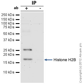 Histone H2B was immunoprecipitated using 0.5mg Hela whole cell extract, 5µg of Mouse monoclonal to Histone H2B and 50µl of protein G magnetic beads (+). No antibody was added to the control (-). The antibody was incubated under agitation with Protein G beads for 10min, Hela whole cell extract lysate diluted in RIPA buffer was added to each sample and incubated for a further 10min under agitation.Proteins were eluted by addition of 40µl SDS loading buffer and incubated for 10min at 70oC; 10µl of each sample was separated on a SDS PAGE gel, transferred to a nitrocellulose membrane, blocked with 5% BSA and probed with ab52484.Secondary: Protein G-HRP at 1/500 dilution.Band: 17kDa; Histone H2B; non specific bands - 26kDa: We are unsure as to the identity of this extra band.
Product References
SILAC-based proteomic profiling of the human MDA-MB-231 metastatic breast cancer - SILAC-based proteomic profiling of the human MDA-MB-231 metastatic breast cancer
Hoedt E, Chaoui K, Huvent I, Mariller C, Monsarrat B, Burlet-Schiltz O, Pierce A. PLoS One. 2014 Aug 12;9(8):e104563.
The tumor suppressor CDC73 interacts with the ring finger proteins RNF20 and - The tumor suppressor CDC73 interacts with the ring finger proteins RNF20 and
Hahn MA, Dickson KA, Jackson S, Clarkson A, Gill AJ, Marsh DJ. Hum Mol Genet. 2012 Feb 1;21(3):559-68.
Induction of gamma-H2AX foci in human exfoliated buccal cells after in vitro - Induction of gamma-H2AX foci in human exfoliated buccal cells after in vitro
Gonzalez JE, Roch-Lefevre SH, Mandina T, Garcia O, Roy L. Int J Radiat Biol. 2010 Sep;86(9):752-9.