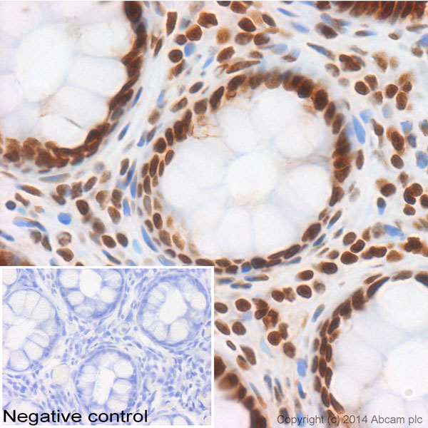
IHC image of ab52985 staining Histone H2B in human colon formalin fixed paraffin embedded tissue sections, performed on a Leica Bond. The section was pre-treated using heat mediated antigen retrieval with sodium citrate buffer (pH6, epitope retrieval solution 1) for 20 mins. The section was then incubated with ab52985, 1/1000 dilution, for 15 mins at room temperature and detected using an HRP conjugated compact polymer system. DAB was used as the chromogen. The section was then counterstained with haematoxylin and mounted with DPX. No primary antibody was used in the negative control (shown on the inset). For other IHC staining systems (automated and non-automated) customers should optimize variable parameters such as antigen retrieval conditions, primary antibody concentration and antibody incubation times.
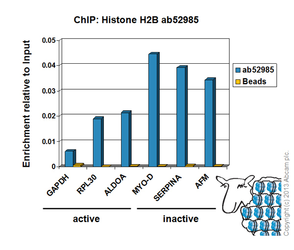
Chromatin was prepared from HeLa cells according to the Abcam X-ChIP protocol. Cells were fixed with formaldehyde for 10 minutes. The ChIP was performed with 25µg of chromatin, 5µg of ab52985 (blue), and 20µl of Protein A/G sepharose beads. No antibody was added to the beads control (yellow). The immunoprecipitated DNA was quantified by real time PCR (Taqman approach). Primers and probes are located in the first kb of the transcribed region.
![Anti-Histone H2B antibody [EP819Y] - ChIP Grade (ab52985) at 1/10000 dilution + A431 cell lysate at 10 µgSecondarygoat anti-rabbit HRP labelled at 1/2000 dilution](http://www.bioprodhub.com/system/product_images/ab_products/2/sub_3/1425_ab52985_WB_1.jpg)
Anti-Histone H2B antibody [EP819Y] - ChIP Grade (ab52985) at 1/10000 dilution + A431 cell lysate at 10 µgSecondarygoat anti-rabbit HRP labelled at 1/2000 dilution

ab52985, at a 1/100 dilution, staining Human Histone H2B in breast carcinoma using Immunohistochemistry, Paraffin Embedded tissue.ab52985, at a 1/100 dilution, staining Human Histone H2B in breast carcinoma using Immunohistochemistry, Paraffin Embedded tissue.