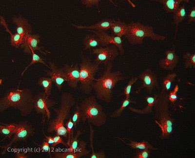
ICC/IF image of ab134211 stained HepG2 cells. The cells were 100% methanol fixed (5 min) and then incubated in 1%BSA / 10% normal goat serum / 0.3M glycine in 0.1% PBS-Tween for 1h to permeabilise the cells and block non-specific protein-protein interactions. The cells were then incubated with the antibody ab134211 at 0.1µg/ml overnight at +4°C. The secondary antibody (green) was DyLight® 488 goat anti- chicken IgY (H+L) used at a 1/1000 dilution for 1h. Alexa Fluor® 594 WGA was used to label plasma membranes (red) at a 1/200 dilution for 1h. DAPI was used to stain the cell nuclei (blue) at a concentration of 1.43µM.
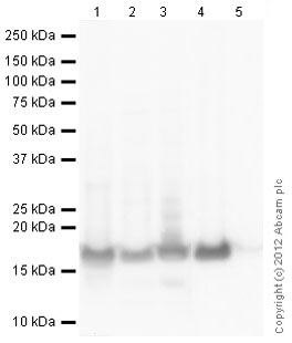
All lanes : Anti-Histone H2B antibody (ab134211) at 1 µg/mlLane 1 : Calf Thymus Histone Preparation Nuclear Lysate at 0.2 µgLane 2 : HeLa (Human epithelial carcinoma cell line) Whole Cell Lysate at 10 µgLane 3 : HeLa Histone Preparation Nuclear Lysate at 2.5 µgLane 4 : Histone H2B Recombinant Protein at 20 µgLane 5 : Histone H3.1 Recombinant Protein at 0.1 µgSecondaryGoat Anti-Chicken IgY H&L (HRP) (ab6877) at 1/3000 dilutiondeveloped using the ECL techniquePerformed under reducing conditions.
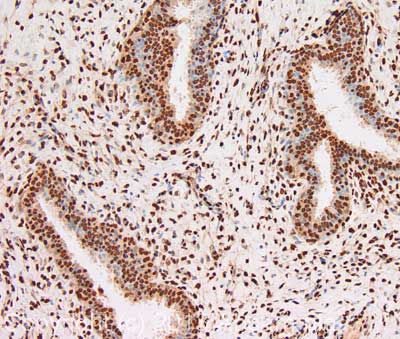
IHC image of Histone H2B staining in human breast fibroadenoma formalin fixed paraffin embedded tissue section, performed on a Leica BondTM system using the standard protocol B. The section was pre-treated using heat mediated antigen retrieval with sodium citrate buffer (pH6, epitope retrieval solution 1) for 20 mins. The section was then incubated with ab134211, 1µg/ml, for 15 mins at room temperature. A goat anti-chicken biotinylated secondary antibody was used to detect the primary, and visualized using an HRP conjugated ABC system. DAB was used as the chromogen. The section was then counterstained with haematoxylin and mounted with DPX. For other IHC staining systems (automated and non-automated) customers should optimize variable parameters such as antigen retrieval conditions, primary antibody concentration and antibody incubation times.