Anti-Histone H2A.X antibody
| Name | Anti-Histone H2A.X antibody |
|---|---|
| Supplier | Abcam |
| Catalog | ab140498 |
| Prices | $384.00 |
| Sizes | 100 µg |
| Host | Goat |
| Clonality | Polyclonal |
| Isotype | IgG |
| Applications | WB IHC-P |
| Species Reactivities | Mouse, Human, Rabbit, Monkey, Ape |
| Antigen | Synthetic peptide corresponding to Human Histone H2A |
| Description | Goat Polyclonal |
| Gene | H2AFX |
| Conjugate | Unconjugated |
| Supplier Page | Shop |
Product images
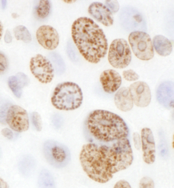 Immunohistochemistry (Formalin/PFA-fixed paraffin-embedded sections) analysis of mouse renal cell carcinoma tissue labelling Histone H2A.X with ab140498 at 1/1000 (1µg/ml). Detection: DAB.
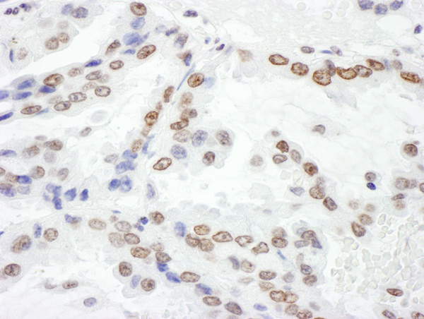 Immunohistochemistry (Formalin/PFA-fixed paraffin-embedded sections) analysis of human lung cancer tissue labelling Histone H2A.X with ab140498 at 1/5000 (0.2µg/ml). Detection: DAB.
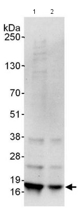 All lanes : Anti-Histone H2A.X antibody (ab140498) at 0.4 µg/mlLane 1 : 293T whole cell lysate at 50 µgLane 2 : 293T whole cell lysate at 15 µgdeveloped using the ECL technique