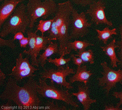
ICC/IF image of ab129217 stained HeLa cells. The cells were exposed to 2000µJ/cm2 of UV, incubated at 37˚C for a further 24 hours. The cells were then 100% methanol fixed (5 min) and then incubated in 1%BSA / 10% normal goat serum / 0.3M glycine in 0.1% PBS-Tween for 1h to permeabilise the cells and block non-specific protein-protein interactions. The cells were then incubated with the antibody (ab129217, 1µg/ml) overnight at +4°C. The secondary antibody (green) was ab96899, DyLight® 488 goat anti-rabbit IgG (H+L) used at a 1/250 dilution for 1h. Alexa Fluor® 594 WGA was used to label plasma membranes (red) at a 1/200 dilution for 1h. DAPI was used to stain the cell nuclei (blue) at a concentration of 1.43µM.
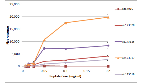
All batches of ab129217 are tested in Peptide Array against peptides to different Histone H2A.X modifications. Six dilutions of each peptide are printed on to the Peptide Array in triplicate and results are averaged before being plotted on to a graph. Results show strong binding to Histone H2A.X - acetyl K5 peptide (ab173317), indicating that this antibody specifically recognises the Histone H2A.X - acetyl K5 modification.ab173317 - Histone H2A.X - acetyl K5ab173318 - Histone H2A.X - unmodifiedab173320 - Histone H2A - unmodifiedab173326 - Histone H2A acethyl K5ab54016 - Histone H2A acetyl K9
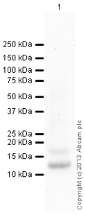
Anti-Histone H2A.X (acetyl K5) antibody (ab129217) at 1 µg/ml + HeLa - Sodium butyrate treated at 10 µgSecondaryDonkey Anti-Rabbit IgG H&L preadsorbed (ab97081) at 1/10000 dilutiondeveloped using the ECL techniquePerformed under reducing conditions.