Phosphatase and Tensin Homolog (
PTEN) is a
tumor suppressor which acts as an antagonist to phosphatidylinositol 3-kinase (PI3K) signaling. PTEN exerts enzymatic activity as a phosphatidylinositol-3,4,5-trisphosphate (PIP3) phosphatase, opposing PI3K activity by reducing availability of PIP3 to proliferating cells. Loss of PTEN function leads to elevated PIP3 and increased activation of PI3K/
AKT signaling in many types of
cancer.
PINK1 (PTEN induced putative kinase 1) protein contains a N-terminal mitochondrial targeting sequence, putative transmembrane helix, linker region, serine (Ser65)/threonine (Thr257) kinase domain and C-terminal segment. PINK1 is translated in the cytosol, then translocated to the outer mitochondrial membrane where it is rapidly cleaved and degraded as a part of normal mitochondrial function. In damaged (depolarized)
mitochondria, PINK1 becomes stabilized and accumulates, resulting in the subsequent phosphorylation of numerous proteins on the mitochondrial surface.
When PINK1 is imported into the cell, mitochondrial processing peptidase, presenilin-associated rhomboid-like protease and
AFG3L2 cleave PINK1 and tag it for the ubiquitin-proteasome pathway, keeping low PINK1 protein expression at basal conditions (1,2). Accumulation of PINK1 in mitochondria indicate damage. PINK1 maintains mitochondrial function/integrity, provides protection against mitochondrial dysfunction during cellular stress, and is involved in the clearance of damaged mitochondria via selective
autophagy (mitophagy) (3). PINK1 has a theoretical molecular weight of 63 kDa and undergoes proteolytic processing to generate at least two cleaved forms (55 kDa and 42 kDa).
Ultimately
PARK2 (E3 Ubiquitin Ligase Parkin) is recruited to the damaged mitochondria where it is activated by 1) PINK-mediated phosphorylation of PARK2 at serine 65, and 2) PARK2 interaction with phosphorylated
ubiquitin (also phosphorylated by PINK1 on serine 65) (4,5). There is a strong interplay between Parkin and PINK1, where loss-of-function of human PINK1 results in mitochondrial pathology and can be rescued by Parkin (2,4,5). Mutations in either Parkin or PINK1 alter mitochondrial turnover, resulting in the accumulation of defective mitochondria and, ultimately,
neurodegeneration in
Parkinson's disease. Mutations in the PINK1 gene located within the PARK6 locus on chromosome 1p35-p36 have been identified in patients with early-onset Parkinson's disease (6).
References
1.Rasool, S., Soya, N., Truong, L., Croteau, N., Lukacs, G. L., & Trempe, J. F. (2018). PINK1 autophosphorylation is required for ubiquitin recognition. EMBO Rep, 19(4). doi:10.15252/embr.201744981
2.Shiba-Fukushima, K., Arano, T., Matsumoto, G., Inoshita, T., Yoshida, S., Ishihama, Y., . . . Imai, Y. (2014). Phosphorylation of mitochondrial polyubiquitin by PINK1 promotes Parkin mitochondrial tethering. PLoS Genet, 10(12), e1004861. doi:10.1371/journal.pgen.1004861
3.Vives-Bauza, C., Zhou, C., Huang, Y., Cui, M., de Vries, R. L., Kim, J., . . . Przedborski, S. (2010). PINK1-dependent recruitment of Parkin to mitochondria in mitophagy. Proc Natl Acad Sci U S A, 107(1), 378-383. doi:10.1073/pnas.0911187107
4.McWilliams, T. G., Barini, E., Pohjolan-Pirhonen, R., Brooks, S. P., Singh, F., Burel, S., . . . Muqit, M. M. K. (2018). Phosphorylation of Parkin at serine 65 is essential for its activation in vivo. Open Biol, 8(11). doi:10.1098/rsob.180108
5.Exner, N., Treske, B., Paquet, D., Holmstrom, K., Schiesling, C., Gispert, S., . . . Haass, C. (2007). Loss-of-function of human PINK1 results in mitochondrial pathology and can be rescued by parkin. J Neurosci, 27(45), 12413-12418. doi:10.1523/jneurosci.0719-07.2007
6.Valente, E. M., Bentivoglio, A. R., Dixon, P. H., Ferraris, A., Ialongo, T., Frontali, M., . . . Wood, N. W. (2001). Localization of a novel locus for autosomal recessive early-onset parkinsonism, PARK6, on human chromosome 1p35-p36. Am J Hum Genet, 68(4), 895-900. doi:10.1086/319522


![Western Blot Parkin Antibody [Unconjugated]](https://images.novusbio.com/images2/Parkin_AF1438_Western_Blot_22174.jpg)
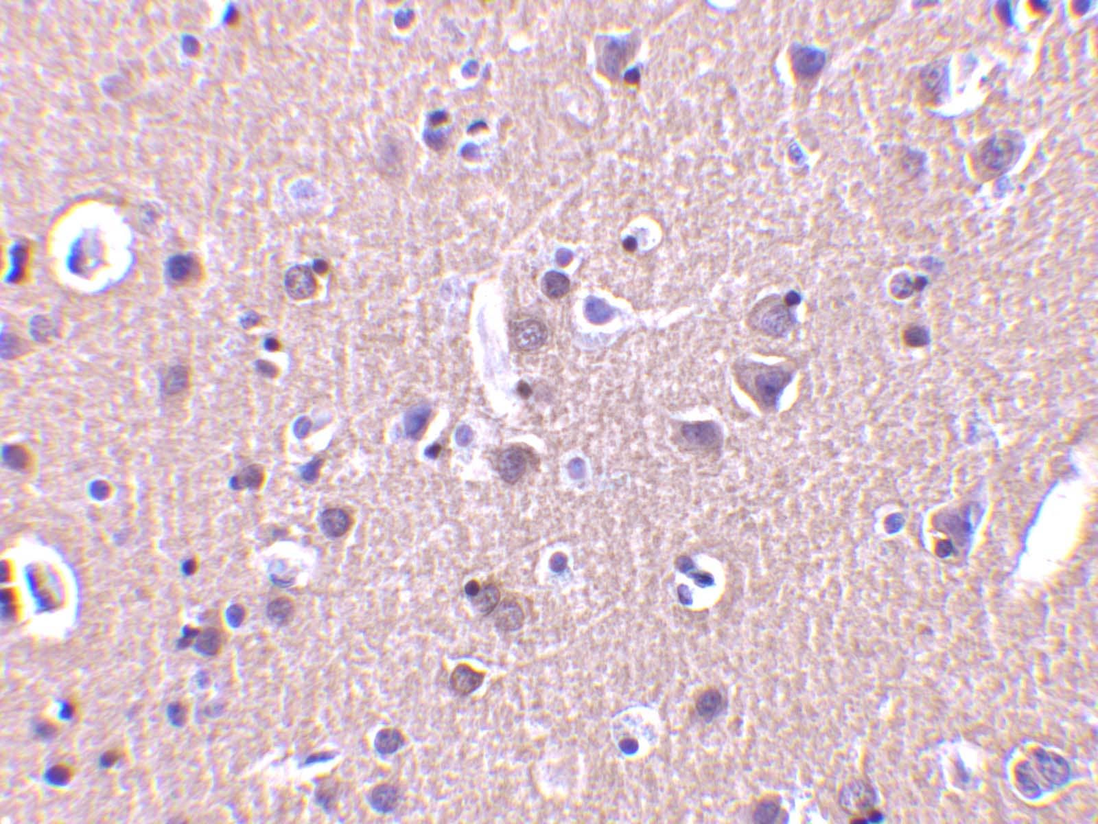
![Immunohistochemistry Parkin Antibody [Unconjugated]](https://images.novusbio.com/images2/Parkin_AF1438_Immunohistochemistry_6700.jpg)


![Western Blot HTRA2/Omi Antibody [Unconjugated]](https://images.novusbio.com/images2/HTRA2_AF1458_Western_Blot_5114.jpg)
![Simple Western HTRA2/Omi Antibody [Unconjugated]](https://images.novusbio.com/images2/16328.jpg)
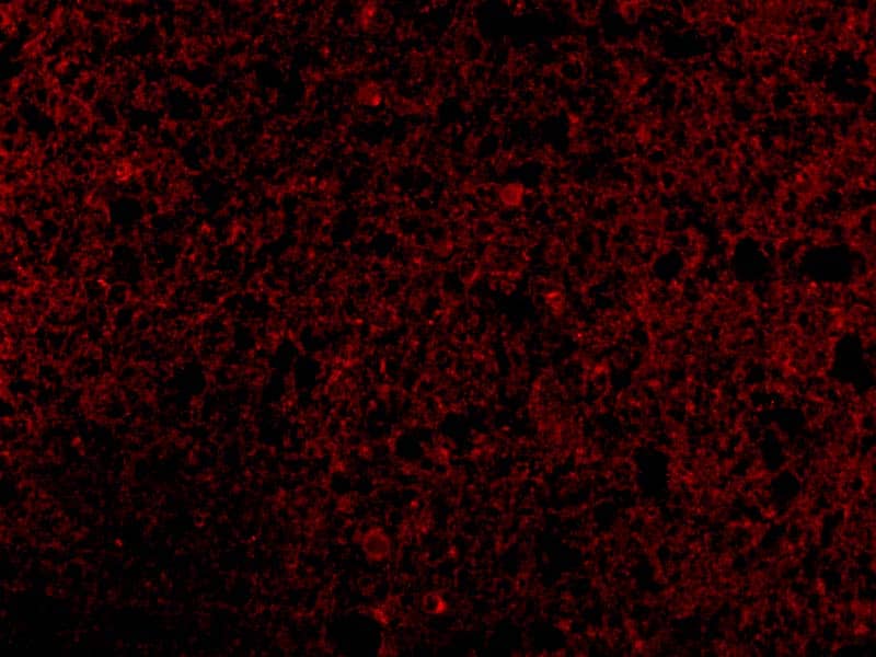
![Immunocytochemistry HTRA2/Omi Antibody [Unconjugated]](https://images.novusbio.com/images2/HTRA2_AF1458_Immunocytochemistry_9924.jpg)


![Western Blot PTEN Antibody [Unconjugated]](https://images.novusbio.com/images2/PTEN_AF847_Western_Blot_5922.jpg)
![Simple Western PTEN Antibody [Unconjugated]](https://images.novusbio.com/images2/16342.jpg)
![Knockout Validated PTEN Antibody [Unconjugated]](https://images.novusbio.com/images2/PTEN_AF847_Knockout_Validated_22996.jpg)


![Simple Western Glucosylceramidase/GBA Antibody (812201) [Unconjugated]](https://images.novusbio.com/images2/16239.jpg)
![Western Blot Glucosylceramidase/GBA Antibody (812201) [Unconjugated]](https://images.novusbio.com/images2/Glucosylceramidase_MAB7410_Western_Blot_12708.jpg)
![Immunocytochemistry Glucosylceramidase/GBA Antibody (812201) [Unconjugated]](https://images.novusbio.com/images2/Glucosylceramidase_MAB7410_Immunocytochemistry_12732.jpg)


