")
ATP5B Antibody (H-3): sc-166443
- ATP5B Antibody (H-3) is a mouse monoclonal IgG2a λ, cited in 7 publications, provided at 200 µg/ml
- specific for an epitope mapping between amino acids 511-530 at the C-terminus of ATP5B of human origin
- recommended for detection of ATP5B of mouse, rat, human and origin by WB, IP, IF, IHC(P) and ELISA; also reactive with additional species, including and equine, canine, bovine, porcine and avian
- m-IgGλ BP-HRP (mouse IgGλ binding protein-HRP) is the preferred secondary detection reagent for ATP5B Antibody (H-3) for WB and IHC(P) applications. This reagent is now offered in a bundle with ATP5B Antibody (H-3) (see ordering information below). For additional m-IgGλ BP conjugates see our complete list of Mouse IgG Binding Proteins.
QUICK LINKS
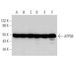
ATP5B Antibody (H-3) is a mouse monoclonal IgG2a antibody that detects ATP5B in mouse, rat, and human samples through various applications including western blotting (WB), immunoprecipitation (IP), immunofluorescence (IF), immunohistochemistry, and enzyme-linked immunosorbent assay (ELISA). ATP5B, also known as ATPMB or ATPSB, is a crucial 529 amino acid protein that plays a vital role in the mitochondrial membrane as a subunit of the F0 complex of ATP synthase, which is essential for ATP production during oxidative phosphorylation. The F0 complex, along with the F1 complex, utilizes the electrochemical gradient across the inner mitochondrial membrane to synthesize ATP, a process that is fundamental for cellular energy metabolism. ATP5B (H-3) antibody recognizes a protein critical for ATP synthesis and overall efficiency of mitochondrial respiration. ATP5B is encoded by a nuclear gene and assembles with other subunits encoded by both mitochondrial and nuclear genes, highlighting the intricate coordination required for mitochondrial function. ATP5B mRNA expression varies significantly among different tissues, with high levels found in the heart, moderate levels in skeletal muscle, and minimal levels in the liver and kidney, indicating its specialized role in energy-demanding tissues. Ets family transcription factors influence ATP5B gene expression, enhancing ATP5B levels in rapidly proliferating cells, further underscoring the importance of this protein in cellular metabolism and energy homeostasis.
Alexa Fluor® is a trademark of Molecular Probes Inc., OR., USA
LI-COR® and Odyssey® are registered trademarks of LI-COR Biosciences
ATP5B Antibody (H-3) References:
- Bacillus anthracis spores influence ATP synthase activity in murine macrophages. | Seo, GM., et al. 2008. J Microbiol Biotechnol. 18: 778-83. PMID: 18467876
- Mitochondrial F1Fo-ATP synthase translocates to cell surface in hepatocytes and has high activity in tumor-like acidic and hypoxic environment. | Ma, Z., et al. 2010. Acta Biochim Biophys Sin (Shanghai). 42: 530-7. PMID: 20705594
- [Effects of electrode on epileptogenic focus potential and expressions of the beta subunit of ATP synthase in rats with penicillin-induced epilepsy]. | He, G., et al. 2012. Sheng Wu Yi Xue Gong Cheng Xue Za Zhi. 29: 287-90. PMID: 22616176
- Potential therapeutic target for malignant paragangliomas: ATP synthase on the surface of paraganglioma cells. | Fliedner, SM., et al. 2015. Am J Cancer Res. 5: 1558-70. PMID: 26101719
- ATP synthase subunit-β down-regulation aggravates diabetic nephropathy. | Guan, SS., et al. 2015. Sci Rep. 5: 14561. PMID: 26449648
- ATP synthase β-subunit abnormality in pancreas islets of rats with polycystic ovary syndrome and type 2 diabetes mellitus. | Li, W., et al. 2017. J Huazhong Univ Sci Technolog Med Sci. 37: 210-216. PMID: 28397049
- Systematic Analysis of the Expression of the Mitochondrial ATP Synthase (Complex V) Subunits in Clear Cell Renal Cell Carcinoma. | Brüggemann, M., et al. 2017. Transl Oncol. 10: 661-668. PMID: 28672194
- FUS interacts with ATP synthase beta subunit and induces mitochondrial unfolded protein response in cellular and animal models. | Deng, J., et al. 2018. Proc Natl Acad Sci U S A. 115: E9678-E9686. PMID: 30249657
- Acquired Resistance to HER2-Targeted Therapies Creates Vulnerability to ATP Synthase Inhibition. | Gale, M., et al. 2020. Cancer Res. 80: 524-535. PMID: 31690671
- ATP synthase subunit ATP5B interacts with TGEV Nsp2 and acts as a negative regulator of TGEV replication. | Wang, Y., et al. 2024. Virulence. 15: 2397492. PMID: 39239724
Ordering Information
| Product Name | Catalog # | UNIT | Price | Qty | FAVORITES | |
ATP5B Antibody (H-3) | sc-166443 | 200 µg/ml | $316.00 | |||
ATP5B Antibody (H-3): m-IgGλ BP-HRP Bundle | sc-521611 | 200 µg Ab, 40 µg BP | $354.00 | |||
ATP5B (H-3) Neutralizing Peptide | sc-166443 P | 100 µg/0.5 ml | $68.00 |