")
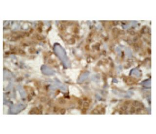
: sc-53617. Immunoperoxidase staining of formalin-fixed, paraffin-embedded human prostate cancer showing cytoplasmic and membrane staining.")
: sc-53617. Western blot analysis of human recombinant CYP3A4 (A), CYP3A7 (B), CYP3A5 (C) and negative control (D) fusion proteins.")
CYP3A7 Antibody (F19 P2 H2): sc-53617
- CYP3A7 Antibody (F19 P2 H2) is a mouse monoclonal IgG1 κ CYP3A7 antibody, cited in 1 publications, provided at 200 µg/ml
- raised against the C-terminus of CYP3A7 of human origin
- CYP3A7 Antibody (F19 P2 H2) is recommended for detection of Cytochrome P4503A7 of human origin by WB, IP, IF and IHC(P)
- Anti-CYP3A7 Antibody (F19 P2 H2) is available conjugated to agarose for IP; HRP for WB, IHC(P) and ELISA; and to either phycoerythrin or FITC for IF, IHC(P) and FCM
- also available conjugated to Alexa Fluor® 488, Alexa Fluor® 546, Alexa Fluor® 594 or Alexa Fluor® 647 for WB (RGB), IF, IHC(P) and FCM, and for use with RGB fluorescent imaging systems, such as iBright™ FL1000, FluorChem™, Typhoon, Azure and other comparable systems
- also available conjugated to Alexa Fluor® 680 or Alexa Fluor® 790 for WB (NIR), IF and FCM; for use with Near-Infrared (NIR) detection systems, such as LI-COR®Odyssey®, iBright™ FL1000, FluorChem™, Typhoon, Azure and other comparable systems
- Contact our Technical Service Department (or your local Distributor) for more information on how to receive a FREE 10 µg sample of CYP3A7 (F19 P2 H2): sc-53617.
- m-IgG Fc BP-HRP and m-IgG1 BP-HRP are the preferred secondary detection reagents for CYP3A7 Antibody (F19 P2 H2) for WB and IHC(P) applications. These reagents are now offered in bundles with CYP3A7 Antibody (F19 P2 H2) (see ordering information below).
CYP3A7 Antibody (F19 P2 H2) is a mouse monoclonal IgG1 kappa light chain antibody that detects CYP3A7 of human origin by western blotting (WB), immunoprecipitation (IP), immunofluorescence (IF), and immunohistochemistry with paraffin-embedded sections (IHCP). Anti-CYP3A7 antibody (F19 P2 H2) is available in both non-conjugated and various conjugated forms, including agarose, horseradish peroxidase (HRP), phycoerythrin (PE), fluorescein isothiocyanate (FITC), and multiple Alexa Fluor® conjugates. CYP3A7 plays a crucial role in drug metabolism, particularly in the biotransformation of various therapeutic agents, which is essential for determining drug efficacy and safety. CYP3A7 is predominantly expressed in the fetal liver and is involved in the metabolism of steroids and other lipids, highlighting its importance in developmental pharmacology. The CYP3A gene family, which includes CYP3A4, CYP3A5, CYP3A7, and CYP3A43, is responsible for the metabolism of over 50% of clinically used drugs, making CYP3A7 a significant player in pharmacogenomics. Understanding CYP3A7 function is vital for predicting individual responses to medications, especially in pediatric populations where expression can differ significantly from adults. The CYP3A gene cluster is located on chromosome 7q22.1, and its members are strategically localized in organs associated with drug disposition, such as the liver, gastrointestinal tract, and kidneys, further emphasizing CYP3A7′s relevance in pharmacological studies.
Alexa Fluor® is a trademark of Molecular Probes Inc., OR., USA
LI-COR® and Odyssey® are registered trademarks of LI-COR Biosciences
CYP3A7 Antibody (F19 P2 H2) References:
- Two linked mutations in transcriptional regulatory elements of the CYP3A5 gene constitute the major genetic determinant of polymorphic activity in humans. | Paulussen, A., et al. 2000. Pharmacogenetics. 10: 415-24. PMID: 10898111
- Feed-forward regulation of bile acid detoxification by CYP3A4: studies in humanized transgenic mice. | Stedman, C., et al. 2004. J Biol Chem. 279: 11336-43. PMID: 14681232
- Crystal structures of human cytochrome P450 3A4 bound to metyrapone and progesterone. | Williams, PA., et al. 2004. Science. 305: 683-6. PMID: 15256616
- Cytochrome p450 profile of colorectal cancer: identification of markers of prognosis. | Kumarakulasingham, M., et al. 2005. Clin Cancer Res. 11: 3758-65. PMID: 15897573
Ordering Information
| Product Name | Catalog # | UNIT | Price | Qty | FAVORITES | |
CYP3A7 Antibody (F19 P2 H2) | sc-53617 | 200 µg/ml | $316.00 | |||
CYP3A7 Antibody (F19 P2 H2): m-IgG Fc BP-HRP Bundle | sc-526836 | 200 µg Ab; 10 µg BP | $354.00 | |||
CYP3A7 Antibody (F19 P2 H2): m-IgG1 BP-HRP Bundle | sc-532209 | 200 µg Ab; 20 µg BP | $354.00 | |||
CYP3A7 Antibody (F19 P2 H2) AC | sc-53617 AC | 500 µg/ml, 25% agarose | $416.00 | |||
CYP3A7 Antibody (F19 P2 H2) HRP | sc-53617 HRP | 200 µg/ml | $316.00 | |||
CYP3A7 Antibody (F19 P2 H2) FITC | sc-53617 FITC | 200 µg/ml | $330.00 | |||
CYP3A7 Antibody (F19 P2 H2) PE | sc-53617 PE | 200 µg/ml | $343.00 | |||
CYP3A7 Antibody (F19 P2 H2) Alexa Fluor® 488 | sc-53617 AF488 | 200 µg/ml | $357.00 | |||
CYP3A7 Antibody (F19 P2 H2) Alexa Fluor® 546 | sc-53617 AF546 | 200 µg/ml | $357.00 | |||
CYP3A7 Antibody (F19 P2 H2) Alexa Fluor® 594 | sc-53617 AF594 | 200 µg/ml | $357.00 | |||
CYP3A7 Antibody (F19 P2 H2) Alexa Fluor® 647 | sc-53617 AF647 | 200 µg/ml | $357.00 | |||
CYP3A7 Antibody (F19 P2 H2) Alexa Fluor® 680 | sc-53617 AF680 | 200 µg/ml | $357.00 | |||
CYP3A7 Antibody (F19 P2 H2) Alexa Fluor® 790 | sc-53617 AF790 | 200 µg/ml | $357.00 |