")
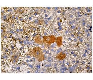
: sc-365943. Immunoperoxidase staining of formalin fixed, paraffin-embedded human adrenal gland tissue showing cytoplasmic staining of subset of glandular cells.")
: sc-365943. Western blot analysis of Ret expression in non-transfected: sc-117752 (A) and human Ret transfected: sc-158925 (B) 293T whole cell lysates.")
Ret Antibody (C-3): sc-365943
- Ret Antibody (C-3) is a mouse monoclonal IgG1 κ Ret antibody, cited in 5 publications, provided at 200 µg/ml
- raised against amino acids 31-330 mapping near the N-terminus of a region conserved between Ret isoforms A and C of human origin
- Ret Antibody (C-3) is recommended for detection of Ret isoforms A and C of human origin by WB, IP, IF, IHC(P) and ELISA
- Anti-Ret Antibody (C-3) is available conjugated to agarose for IP; HRP for WB, IHC(P) and ELISA; and to either phycoerythrin or FITC for IF, IHC(P) and FCM
- also available conjugated to Alexa Fluor® 488, Alexa Fluor® 546, Alexa Fluor® 594 or Alexa Fluor® 647 for WB (RGB), IF, IHC(P) and FCM, and for use with RGB fluorescent imaging systems, such as iBright™ FL1000, FluorChem™, Typhoon, Azure and other comparable systems
- also available conjugated to Alexa Fluor® 680 or Alexa Fluor® 790 for WB (NIR), IF and FCM; for use with Near-Infrared (NIR) detection systems, such as LI-COR®Odyssey®, iBright™ FL1000, FluorChem™, Typhoon, Azure and other comparable systems
- Contact our Technical Service Department (or your local Distributor) for more information on how to receive a FREE 10 µg sample of Ret (C-3): sc-365943.
- m-IgG Fc BP-HRP, m-IgG1 BP-HRP and m-IgGκ BP-HRP are the preferred secondary detection reagents for Ret Antibody (C-3) for WB and IHC(P) applications. These reagents are now offered in bundles with Ret Antibody (C-3) (see ordering information below).
QUICK LINKS
SEE ALSO...
Ret Antibody (C-3) is a mouse monoclonal IgG1 kappa light chain antibody that detects Ret protein of human origin by western blotting (WB), immunoprecipitation (IP), immunofluorescence (IF), immunohistochemistry with paraffin-embedded sections (IHCP), and enzyme-linked immunosorbent assay (ELISA). Ret Antibody (C-3) is available in both non-conjugated and various conjugated forms, including agarose, horseradish peroxidase (HRP), phycoerythrin (PE), fluorescein isothiocyanate (FITC), and multiple Alexa Fluor® conjugates. The Ret proto-oncogene plays a crucial role in cell signaling, particularly in the glial cell line-derived neurotrophic factor (GDNF) signaling pathway, which is essential for the development and maintenance of the nervous system. Ret protein is primarily located on the cell membrane, where Ret (C-3) antibody functions as a receptor tyrosine kinase, facilitating communication between cells and their environment. This localization is vital for mediating cellular responses to growth factors, influencing processes such as cell differentiation, survival, and proliferation. Notably, aberrations in the Ret gene, including tumor-specific rearrangements, have been implicated in various cancers, particularly papillary thyroid carcinoma, where anti-Ret antibody (C-3) leads to the production of fusion proteins that exhibit constitutive activation of the Ret signaling pathway. Such alterations underscore Ret protein′s importance in both normal physiology and disease, making Ret (C-3) monoclonal antibody an invaluable tool for researchers studying its function and implications in cancer biology.
Alexa Fluor® is a trademark of Molecular Probes Inc., OR., USA
LI-COR® and Odyssey® are registered trademarks of LI-COR Biosciences
Ret Antibody (C-3) References:
- Identification of the product of two oncogenic rearranged forms of the RET proto-oncogene in papillary thyroid carcinomas. | Lanzi, C., et al. 1992. Oncogene. 7: 2189-94. PMID: 1437145
- Identification of the ret proto-oncogene products in neuroblastoma and leukemia cells. | Takahashi, M., et al. 1991. Oncogene. 6: 297-301. PMID: 2000222
- Characterization of ret proto-oncogene mRNAs encoding two isoforms of the protein product in a human neuroblastoma cell line. | Tahira, T., et al. 1990. Oncogene. 5: 97-102. PMID: 2181380
- PTC is a novel rearranged form of the ret proto-oncogene and is frequently detected in vivo in human thyroid papillary carcinomas. | Grieco, M., et al. 1990. Cell. 60: 557-63. PMID: 2406025
- Crosstalk of parkin and Ret in dopaminergic neurons. | Kramer, ER. 2015. Oncotarget. 6: 15704-5. PMID: 26202959
- Cloning and expression of the ret proto-oncogene encoding a tyrosine kinase with two potential transmembrane domains. | Takahashi, M., et al. 1988. Oncogene. 3: 571-8. PMID: 3078962
- Targeted therapies for RET-fusion cancer: Dilemmas and breakthrough. | Ding, S., et al. 2020. Biomed Pharmacother. 132: 110901. PMID: 33125973
- Hallmarks of RET and Co-occuring Genomic Alterations in RET-aberrant Cancers. | Adashek, JJ., et al. 2021. Mol Cancer Ther. 20: 1769-1776. PMID: 34493590
- The evolution of RET inhibitor resistance in RET-driven lung and thyroid cancers. | Rosen, EY., et al. 2022. Nat Commun. 13: 1450. PMID: 35304457
- RET is a sex-biased regulator of intestinal tumorigenesis. | Koester, ST., et al. 2023. Front Gastroenterol (Lausanne). 2: PMID: 39148929
- Functional receptor for GDNF encoded by the c-ret proto-oncogene. | Trupp, M., et al. 1996. Nature. 381: 785-9. PMID: 8657281
- RET mutations in human disease. | Pasini, B., et al. 1996. Trends Genet. 12: 138-44. PMID: 8901418
Ordering Information
| Product Name | Catalog # | UNIT | Price | Qty | FAVORITES | |
Ret Antibody (C-3) | sc-365943 | 200 µg/ml | $316.00 | |||
Ret Antibody (C-3): m-IgG Fc BP-HRP Bundle | sc-529522 | 200 µg Ab; 10 µg BP | $354.00 | |||
Ret Antibody (C-3): m-IgGκ BP-HRP Bundle | sc-522436 | 200 µg Ab, 40 µg BP | $354.00 | |||
Ret Antibody (C-3): m-IgG1 BP-HRP Bundle | sc-543559 | 200 µg Ab; 20 µg BP | $354.00 | |||
Ret Antibody (C-3) AC | sc-365943 AC | 500 µg/ml, 25% agarose | $416.00 | |||
Ret Antibody (C-3) HRP | sc-365943 HRP | 200 µg/ml | $316.00 | |||
Ret Antibody (C-3) FITC | sc-365943 FITC | 200 µg/ml | $330.00 | |||
Ret Antibody (C-3) PE | sc-365943 PE | 200 µg/ml | $343.00 | |||
Ret Antibody (C-3) Alexa Fluor® 488 | sc-365943 AF488 | 200 µg/ml | $357.00 | |||
Ret Antibody (C-3) Alexa Fluor® 546 | sc-365943 AF546 | 200 µg/ml | $357.00 | |||
Ret Antibody (C-3) Alexa Fluor® 594 | sc-365943 AF594 | 200 µg/ml | $357.00 | |||
Ret Antibody (C-3) Alexa Fluor® 647 | sc-365943 AF647 | 200 µg/ml | $357.00 | |||
Ret Antibody (C-3) Alexa Fluor® 680 | sc-365943 AF680 | 200 µg/ml | $357.00 | |||
Ret Antibody (C-3) Alexa Fluor® 790 | sc-365943 AF790 | 200 µg/ml | $357.00 |