EGFR protein (Fc Chimera Active)
| Name | EGFR protein (Fc Chimera Active) |
|---|---|
| Supplier | Abcam |
| Catalog | ab84002 |
| Category | Protein |
| Prices | $315.00 |
| Sizes | 25 µg |
| Applications | SDS-PAGE FA |
| Species Reactivities | Human |
| Nature | Recombinant |
| Source | HEK 293 cells |
| Purity | > 95 % by SDS-PAGE. |
| Bioactivity | The ED 50 of ab84002 is typically 60-100 ng/ml as measured by its ability to neutralize EGF mediated proliferation of murine NIH3T3 fibroblasts. |
| SwissProt/Accession | P00533 |
| Gene | EGFR |
| Sequence | Theoretical sequence: LEEKKVCQGTSNKLTQLGTFEDHFLSLQRMFNNCEVVLG NLEITYVQR NYDLSFLKTIQEVAGYVLIALNTVERIPLENLQIIRGNM YYENSYALA VLSNYDANKTGLKELPMRNLQEILHGAVRFSNNPALCNV ESIQWRDIV SSDFLSNMSMDFQNHLGSCQKCDPSCPNGSCWGAGEENC QKLTKIICA QQCSGRCRGKSPSDCCHNQCAAGCTGPRESDCLVCRKFR DEATCKDTC PPLMLYNPTTYQMDVNPEGKYSFGATCVKKCPRNYVVTD HGSCVRACG ADSYEMEEDGVRKCKKCEGPCRKVCNGIGIGEFKDSLSI NATNIKHFK NCTSISGDLHILPVAFRGDSFTHTPPLDPQELDILKTVK EITGFLLIQ AWPENRTDLHAFENLEIIRGRTKQHGQFSLAVVSLNITS LGLRSLKEI SDGDVIISGNKNLCYANTINWKKLFGTSGQKTKIISNRG ENSCKATGQ VCHALCSPEGCWGPEPRDCVSRSSNTKVDKKVEPKSCDK THTCPPCPA PELLGGPSVFLFPPKPKDTLMISRTPEVTCVVVDVSHED PEVKFNWYV DGVEVHNAKTKPREEQYNSTYRVVSVLTVLHQDWLNGKE YKCKVSNKA LPAPIEKTISKAKGQPREPQVYTLPPSRDELTKNQVSLT CLVKGFYPS DIAVEWESNGQPENNYKTTPPVLDSDGSFFLYSKLTVDK SRWQQGNVF SCSVMHEALHNHYTQKSLSLSPGK |
| Supplier Page | Shop |
Product images
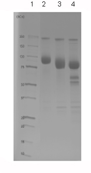 Lane 1 – MW markers; Lane 2 – ab84002; Lane 3 – ab84002 treated with PNGase F to remove potential N-linked glycans; Lane 4 – ab84002 treated with a glycosidase cocktail to remove potential N and O-linked glycans. Approximately 5 µg of protein was loaded per lane; Gel was stained using Deep Purple™. Drop in MW after treatment with PNGase F indicates presence of N-linked glycans. A further drop in MW after treatment with the glycosidase cocktail indicates the presence of O-linked glycans. Additional bands in lane 3 and lane 4 are glycosidase enzymes.
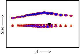 Post-translational modifications result in protein heterogeneity. The densitometry scan demonstrates the purified human cell expressed protein exists in multiple glycoforms, which differ according to their level of post-translational modification. The triangle indicates theoretical pI and MW of the protein.