![Standard curve for Transferrin (Analyte: ab83560 ); dilution range 1pg/ml to 1µg/ml using Capture Antibody Mouse monoclonal [HTF-14] to Transferrin (ab769) at 1µg/ml and Detector Antibody Rabbit polyclonal to Transferrin (ab9538) at 0.5µg/ml.](http://www.bioprodhub.com/system/product_images/pp_products/2/sub_1/2104_Transferrin-Proteins-and-Peptides-ab83560-6.jpg)
Standard curve for Transferrin (Analyte: ab83560 ); dilution range 1pg/ml to 1µg/ml using Capture Antibody Mouse monoclonal [HTF-14] to Transferrin (ab769) at 1µg/ml and Detector Antibody Rabbit polyclonal to Transferrin (ab9538) at 0.5µg/ml.

Lane 1 – MW markers; Lane 2 – ab83560; Lane 3 – ab83560 treated with PNGase F to remove potential N-linked glycans; Lane 4 – ab83560 treated with a glycosidase cocktail to remove potential N- and Olinked glycans. Approximately 5 µg of protein was loaded per lane; Gel was stained using Coomassie. Drop in MW after treatment with PNGase F indicates presence of N-linked glycans. A further drop in MW after treatment with the glycosidase cocktail indicates the presence of O-linked glycans. Additional bands in lane 3 and lane 4 are glycosidase enzymes.

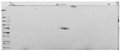
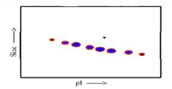
A sample of ab83560 without carrier protein was reduced and alkylated and focused on a 3-10 IPG strip then run on a 4-20% Tris HCl 2D gel. Approximately 40 µg of protein was load; Gel was stained using Deep Purple™. Spot train indicates presence of multiple isoforms of Transferrin. Spots within the spot train were cut from the gel and identified as Transferrin by protein mass fingerprinting.

Post-translational modifications result in protein heterogeneity. The densitometry scan demonstrates that ab83560 exists in multiple isoforms, which differ according to their level of posttranslational modification. Expression of these isoforms is highly significant for cell biology, as they more closely resemble the native human proteins. The triangle indicates theoretical pI and MW of the protein.
![Standard curve for Transferrin (Analyte: ab83560 ); dilution range 1pg/ml to 1µg/ml using Capture Antibody Mouse monoclonal [HTF-14] to Transferrin (ab769) at 1µg/ml and Detector Antibody Rabbit polyclonal to Transferrin (ab9538) at 0.5µg/ml.](http://www.bioprodhub.com/system/product_images/pp_products/2/sub_1/2104_Transferrin-Proteins-and-Peptides-ab83560-6.jpg)


