PathScan ® Phospho-FLT3 (panTyr) Sandwich ELISA Kit
| Name | PathScan ® Phospho-FLT3 (panTyr) Sandwich ELISA Kit |
|---|---|
| Supplier | Cell Signaling Technology |
| Catalog | 7761 |
| Category | ELISA Kit |
| Prices | $489.00 |
| Sizes | 1 kit (96 assays) |
| Assay Type | Sandwich |
| Applications | ELISA |
| Species Reactivities | Human |
| Description | CST's PathScan ® Phospho-FLT3 (panTyr) Sandwich ELISA Kit is a solid phase sandwich enzyme-linked immunosorbent assay (ELISA) that detects endogenous levels of tyrosine-phosphorylated FLT3 protein |
| Gene | FLT3 |
| Supplier Page | Shop |
Product images
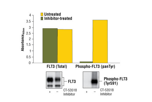 Figure 1: Constitutive phosphorylated of FLT3 in SEM cells is detected by PathScan ® Phospho-FLT3 (panTyr) Sandwich ELISA kit #7761. In contrast, only a low level of phospho-FLT3 protein is detected in SEM cells treated with CT-53518, an inhibitor of FLT3 tyrosine phosphorylation. The inhibitor does not affect the level of total FLT3 protein detected by PathScan ® Total FLT3 Sandwich ELISA kit #7202. Absorbance at 450 nm is shown in the top figure, while the corresponding western blots using Phospho-FLT3 (Tyr591) Antibody #3461 (right panel) or FLT3 Rabbit mAb #3462 (left panel), is shown in the bottom figure.
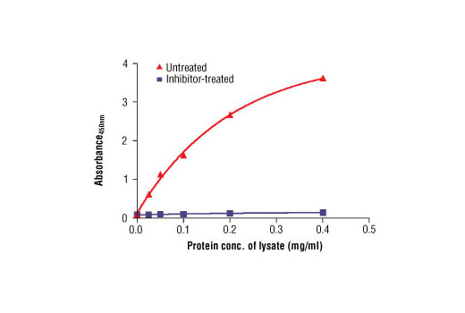 Figure 2: The relationship between total protein concentration of the lysate from untreated or inhibitor-treated SEM cells and absorbance at 450 nm is shown. Unstarved SEM cells (10-6 cells/ml) were left untreated or treated with CT-53518 (50 ng/ml) for 120 min and then lysed.