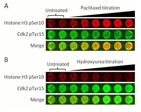
HeLa cells were treated for 24h with varying concentrations of paclitaxel (0.26 – 2000 µM). Histone H3 pSer10 (red) intensity increases with increasing paclitaxel whereas Cdk2 pTyr15 (green) intensity decreases. This is the expected result for paclitaxel treatment: mitotic arrest. (B) HeLa cells were treated for 24h with varying concentrations of hydroxyurea (0.002 – 5 mM). Histone H3 pSer10 (red) intensity decreases with increasing paclitaxel whereas Cdk2 pTyr15 (green) intensity increases. This is the expected result for hydroxyurea treatment: G1/S-phase arrest.

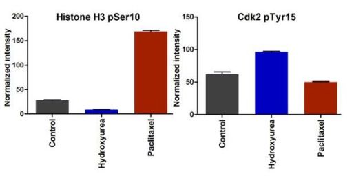
Quantification of the data shown in Image 1. Data shown is for 24 hour treatment with 1 mM hydroxyurea, 333 nM paclitaxel and untreated (Control).

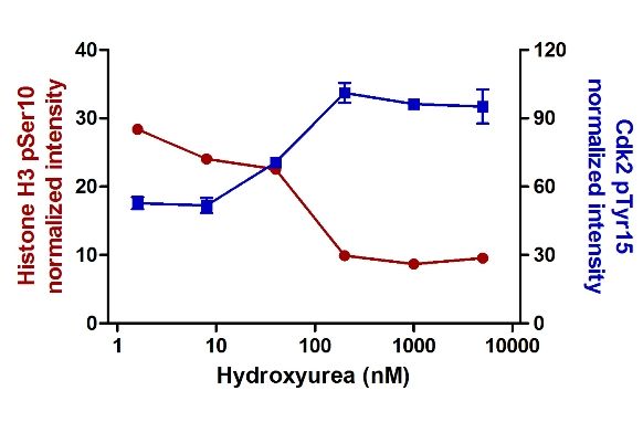
Cdk2 pTyr15 intensity increases with Hydroxyurea treatment dose whereas Histone H3 pSer10 intensity decreases.

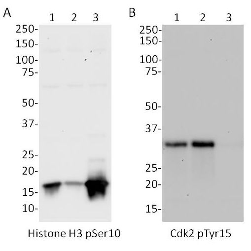
Whole cell lysates from HeLa cells were analyzed by western blot with the primary antibodies used in this assay kit. (A) Histone H3 pSer10 antibody: untreated (lane 1), hydroxyurea = G1/S arrest (lane 2), paclitaxel = G2/M arrest (lane 3). (B) Cdk2 pTyr15 antibody: untreated (lane 1), thymidine = G1/S arrest (lane 2), nocodazole = G2/M arrest (lane 3).