Catalase Human SimpleStep ELISA™ Kit
| Name | Catalase Human SimpleStep ELISA™ Kit |
|---|---|
| Supplier | Abcam |
| Catalog | ab171572 |
| Category | ELISA Kit |
| Prices | $484.00 |
| Sizes | 1 x 96 tests |
| Assay Type | Sandwich (quantitative) |
| Sample Type | Cell culture extracts, Tissue Extracts |
| Detection | Colorimetric |
| Sensitivity | 43 pg/ml |
| Range | 0.31 ng/ml - 20 ng/ml |
| Format | Microplate |
| Applications | ELISA |
| Species Reactivities | Human |
| Gene | CAT |
| Supplier Page | Shop |
Product images
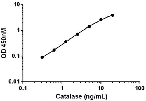
 Background subtracted data from duplicate measurements are plotted.
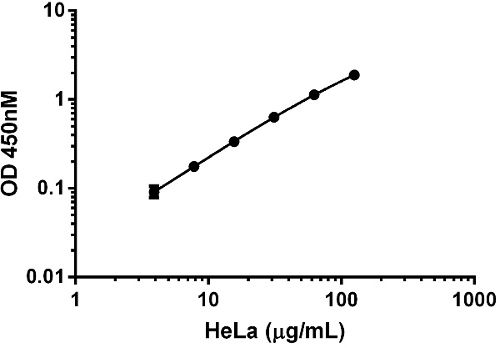
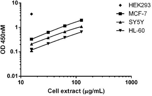 Background subtracted data from duplicate measurements are plotted. Due to relative high expression levels of catalase in the cell line HEK 293, signal saturates when lysate is loaded at > 16 µg/mL.
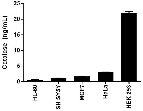 Interpolated values of catalase are plotted for the indicated cell lines based on an extract load of 16 µg/mL.
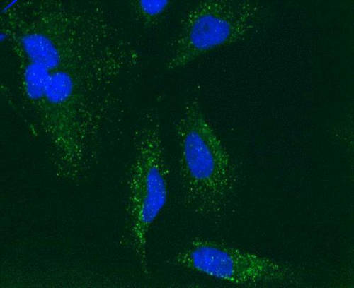 Immunocytochemical staining of Human HeLa cells using the catalase detector antibody in this kit (ab110292). The target protein locates mainly in peroxisomes.