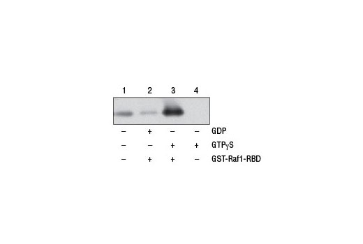
Figure 1. NIH/3T3 cell lysates (500 µl at 1 mg/ml) were treated in vitro with GTPγS or GDP to activate or inactivate Ras (refer to optional step C in protocol). The lysates were then incubated with glutathione resin and GST-Raf1-RBD (lanes 2 and 3). GTPγS-treated lysate was also incubated without GST-Raf1-RBD in the presence of glutathione resin as a negative control (lane 4). Western blot analysis of cell lysate (20 µg, lane 1) or 20 µl of the eluted samples (lanes 2, 3, and 4) was performed using a Ras mouse mAb. Anti-mouse IgG, HRP-linked Antibody #7076 was used as the secondary antibody.

Figure 2. The GTP-bound GTPase pull-down process can be divided into 3 steps as shown. Step 1: Mix sample, binding protein, and glutathione resin in the spin cup and incubate at 4ºC to allow GTP-bound GTPase binding to the glutathione resin through GST-linked binding protein. Step 2: Remove unbound proteins by centrifugation. Step 3: Elute glutathione resin-bound GTPase with SDS buffer. The eluted sample can then be analyzed by western blot.