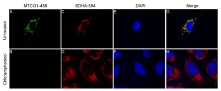
Cells were treated with 10 µM chloramphenicol for a period of 6 days. Results of immunostaining of HeLa cells showing MTCO1 Alexa®488 antibody (ab154477, green) on untreated (A) and chloramphenicol treated (B). SDHA Alexa®594 antibody (red) on untreated (C) and chloramphenicol treated (D). DAPI staining of nucleus (blue) on untreated (E) and chloramphenicol treated (F). Merge of color channels for untreated (G) and chloramphenicol treated (H) cells.

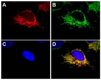
Immunocytochemistry with HeLa cells (100x). A) HeLa stained with anti-SDHA Alexa® 594 antibody (1.0 µg/mL). B) HeLa stained with Anti-HSP60 (1/1000, ab46798), Secondary antibody used was goat anti-rabbit Alexa® 488 (1/1000, ab150077). C) DAPI as nuclear stain (1/10000). D) Merge of color channels to show specificity of signal to mitochondria.

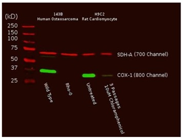
A total of 10 µg from Human or rat cultured cells was probed with the primary and secondary antibodies and scanned with a LI-COR® Odyssey® imager. The two mitochondrial proteins targeted by the two primary antibodies were labeled and visualized specifically despite the presence of thousands of other proteins. Furthermore, reduction of mtDNA levels in human Rho0 (mtDNA-depleted) cells, or inhibition of mitochondrial protein translation by chloramphenicol in rat cells result in specific reduction of COX-I protein while nuclear DNA-encoded SDH-A is unaffected.

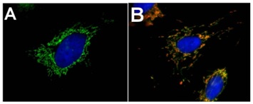
A) Staining of HeLa cells with Immunocytochemistry with HeLa cells (100x) were stained with anti-MTCO1 Alexa® 488 antibody (1.0 µg/mL, ab154477) in green and DAPI in blue, as a nuclear stain. B) Immunocytochemistry with HDFn (100x) cells were stained with Anti-MTOC1 Alexa® 488 antibody (1.0 µg/mL, ab154477) in green, Anti-HSP60 (1/1000, ab46798) as red, and DAPI in blue, as a nuclear stain. Secondary antibody used was goat anti-rabbit dyelight-594 (1/1000, ab96897).


