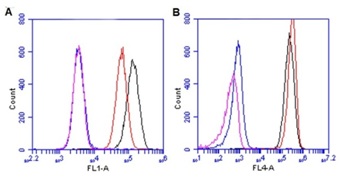
A total of 10,000 gated events were captured for analysis of HeLa cells with or without chloramphenicol (16 µM, 6-days). The results show MTCO1 Alexa®488, FL1(A) and SDHA Alexa®647, FL4(B) co staining for untreated, no anitobdies (blue); untreated with antibodies (black); chloramphenicol, no antibodies (purple); and chloramphenicol with antibodies (red).

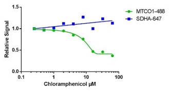
HeLa cells were treated with 2-fold serial dilution of chloramphenicol for 6 days. The relative levels of the mean FL-1 fluorescence (MTCO1 Alexa® 488) and FL-4 fluorescence (SDHA Alexa® 647); a total of 10,000 events were collected.

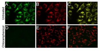
Cells were treated with 30 µM chloramphenicol for a period fo 6 days. Results of immunostaining of HeLa cells showing MTCO1 Alexa® 488 antibody (ab154477, green) on untreated (A) and chloramphenicol treated (D). SDHA Alexa® 647 antibody (ab168536, red) on untreated (B) and chloramphenicol treated (E). Merge of color channels for untreated (C) and chloramphenicol treated (F) cells.

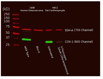
A total of 10 µg from Human or rat cultured cells were probed with the primary and secondary antibodies and scanned with a LI-COR® Odyssey® imager. Reduction of mtDNA levels in human Rho0 (mtDNA depleted) cells, or inhibition of mitochondrial protein translation by chloramphenicol in rat cells result in specific reduction of COX-I protein while nuclear DNA-encoded SDH-A is unaffected.


