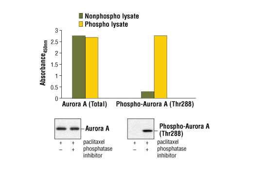
Figure 1. Induced phosphorylation of Aurora A in paclitaxel-treated HeLa cells lysed in the presence of phosphatase inhibitors (phospho lysate) is detected by PathScan® Phospho-Aurora A (Thr288) Sandwich ELISA Kit #7114 (upper, right). In contrast, a low level of phospho-Aurora A protein is detected in paclitaxel-treated HeLa cells lysed without addition of phosphatase inhibitors to the lysis buffer (nonphospho lysate). Similar levels of Aurora A protein from either nonphospho or phospho lysates are detected by PathScan® Total Aurora A Sandwich ELISA Kit #7116 (upper, left). The absorbance readings at 450 nm are shown in the top figure, while the corresponding Western blots using Aurora A Antibody #3092 (left panel) or Phospho-Aurora A (Thr288) Rabbit mAb #3079 (right panel) are shown in the bottom figure.
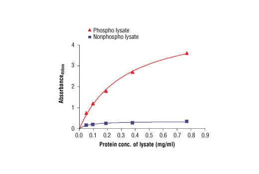
Figure 2: The relationship between protein concentration of phospho or nonphospho Aurora A lysates and the absorbance at 450 nm is shown. Unstarved HeLa cells (85% confluence) treated with paclitaxel (100 nM) for 20 hours were harvested and then lysed in the absence or presence of phosphatase inhibitor.