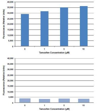
HepG2 cells were seeded in a 96-well plate at a density of 5 x 10⁴ cells/well in EMEM culture medium and incubated O/N at 37°C. The next day, cells were treated with increasing concentrations of tamoxifen and incubated O/N. On the third day, cells were stained with PI and MDC (as described in the assay protocol) and fluorescence was quantified using a plate reader. Top panel: Tamoxifen treatment increases MDC fluorescence intensity, indicating that Tamoxifen treatment leads to an increase in autophagy in HepG2 cells. Bottom panel: Tamoxifen treatment does not cause an increase in PI staining, indicating that at the concentrations used in this experiment, tamoxifen does cause cytotoxicity in HepG2 cells.

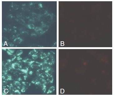
HepG2 cells were seeded at a density of 5 x 10⁴ cells/well and incubated O/N at 37°C. The next day, cells were treated with either vehicle (panel A & B) or 10 µM of tamoxifen for 24 hours. On the third day, cells were stained with PI and MDC as described in the assay protocol. Panel A: MDC staining of HepG2 cells treated with vehicle. There is a basal level of autophagy, indicated by faint silver dot staining of autophagic vacuoles. Panel B: PI staining of HepG2 cells treated with vehicle. There are few dead cells with only background staining of propidium iodide. Panel C: MDC staining of HepG2 cells treated with 10 μM Tamoxifen. There is a clear increase in fluorescence intensity and number of autophagic vacuoles compared to the control cells treated with vehicle. Panel D: PI staining of HepG2 cells treated with 10 μM Tamoxifen, shows similar staining pattern to that of cells treated with vehicle.

