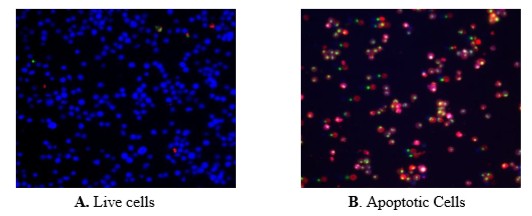
The fluorescence image shows cells that are live (blue, stained by CytoCalcein Violet 450), apoptotic (red, stained by Apopxin Deep Red Indicator), and necrotic (green, indicated by Nuclear Green DCS1staining) in Jurkat cells induced by 1μM staurosporine for 3 hours. The fluorescence images of the cells were taken with a fluorescence microscope through the Violet, Cy5 and FITC channel respectively. Individual images taken from each channel from the same cell population were merged as shown above. A: Non-induced control cells; B: Triple staining of staurosporine-induced cells.
