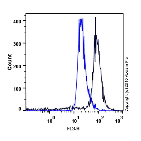
The increase in the fluorescence intensity of the Aggresome Detection Reagent with the addition of MG-132 in Jurkat cells. Jurkat cells were mock induced with 0.08% DMSO (blue line) or induced with 8 μM MG-132 (black line) in a 37ºC, 5% CO2 incubator for 24 hours.
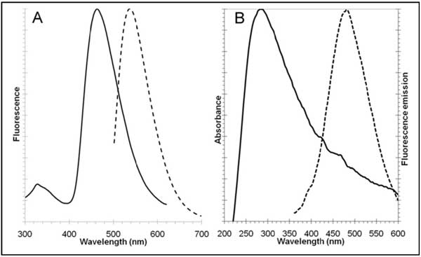
Excitation and fluorescence emission spectra for Aggresome reagent dye, Ex/Em=500/600 nm (panel A, left) and Hoechst 33342, Ex/Em=350/461 nm (panel B, right). All spectra were determined in 1X Assay Buffer.
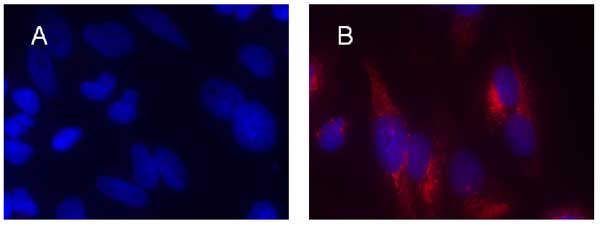
HeLa cells were mock-induced with 0.2% DMSO (panel A) or induced with 5 µM MG-132 (panel B) for 12 hours at 37°C. After treatment, cells were incubated with Aggresome Detection Reagent for 30 minutes.
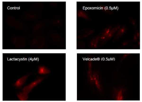
Aggresomes were detected in HeLa cells after overnight incubation with a range of proteasome inhibitors, as observed by fluorescence microscopy.

HeLa cells were pre-treated for 12 hours with 5µM MG-132. Aggresomes detected with Aggresome Detection Reagent (A), showing co-localization with fluorescein-p62 antibody (B), composite image (C), as observed by fluorescence microscopy.
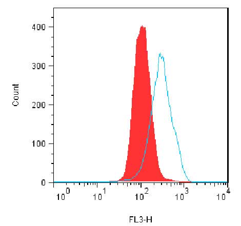
Jurkat cells were mock induced with 0.2% DMSO (block red histogram) or induced with 5 µM MG-132 overnight at 37°C (blue line histogram). After treatment, cells were fixed and incubated with Aggresome Detection Reagent, then analyzed by flow cytometry without washing using a 488 nm laser with fluorescence detection in the FL3 channel. Results are presented as histogram overlays. In MG-132 treated cells, fluorescent green signal increases over 2-fold.


