
Using the S6 RP assay kits or Western blot, S6 RP phosphorylation at Ser240/244 is detected in insulin-treated MCF7 cells (+), compared with MCF7 cells treated with a combination of rapamycin, UCN-01 and U0126 (-), whereas no change in total S6 RP levels was observed.
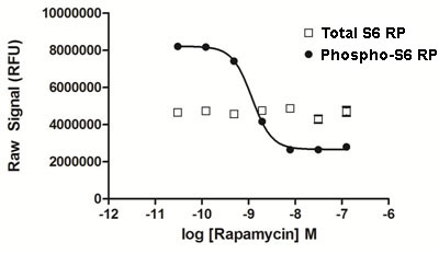
MCF7 cells were seeded at 40K cells/well in medium containing 10% FBS in a 96 well microplate, and cultured overnight. The following day the culture medium was removed, and the cells were treated with various concentrations of rapamycin diluted in serum-free medium for 60 mins. The medium was removed from the wells, and the cells were lysed with 120 µL/well Lysis Mix, with shaking for 10 min. 50 µL of lysate was transferred to replicate wells of a PhosphoTracer assay plate, and Antibody Mix specific for either phospho S6 RP or total S6 RP (50 µL/well) was added to the lysates. The plates were incubated for 1 hr at room temp, with shaking. Plates were washed and Substrate Mix was added. The plates were covered in foil and incubated for 10 min with shaking. Signal in the wells was determined using a plate reader.